Nikon Instruments Inc. | Americas
- en Change Region
- Global Site
Cytogenetics is the study of chromosomes, particularly in relation to diseases caused by chromosome abnormalities. Studies are usually carried out in white blood cells, amniotic fluid, or tissue samples. Chromosomes are only visible in dividing cells and particularly during prophase and metaphase when chromatin condenses. When stained with Giemsa or quinacrine, chromosomes show dark and light banding patterns. Dark bands are generally known as G- or Q- positive bands and light bands as G- or Q-negative bands. G-bands may also be referred to as R-bands because banding patterns reverse under some staining regimens. Other banding patterns can be identified in localised regions of the chromosome, such as close to the centromere.
Banding patterns allow different chromosomes to be identified and paired and provide a way of recognizing chromosome duplications, breaks, deletions, inversions, and translocations - all of which may result in altered phenotypes and disease. Some chromosomal aberrations have been linked to inherited genetic disorders and cancer. The term karyotype is used to describe images of a cell's chromosomes arranged in decreasing size with short arms (p for petit) at the top, and long arms (q for queue or tail) at the bottom.
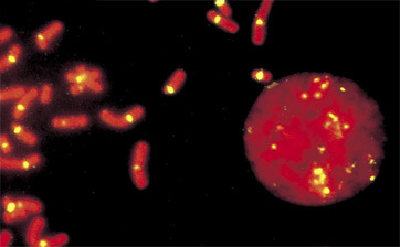
Other important cytogenic techniques include FISH, mFISH, and CGH. FISH has the advantage that it can be carried out in interphase cells and can be carried out in fixed and embedded tissues. FISH can also be used to detect chromosome abnormalities that are beyond the resolving capability of chromosome banding studies. CGH can be used to screen tumor cells for DNA gains and losses that include mutations at chromosomal and subchromosomal levels. Key microscopy techniques in cytogenetics include brightfield and fluorescence microscopy.