Nikon Instruments Inc. | Americas
- en Change Region
- Global Site
Pathology is the study of disease and, in particular, the structural and functional changes that take place in cells and tissues during disease. Knowledge of these changes may be used to diagnose disease, ascertain the cause of death, and to better understand the causes, mechanisms, and consequences of disease.
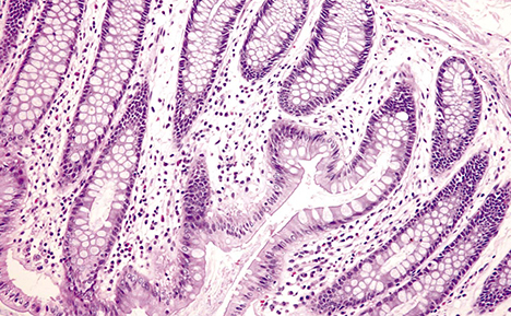
A pathologist may make macroscopic examinations of tissues removed during surgery or after death, examine tissue slices (frozen or fixed) under the microscope, examine exfoliated cells (cytology), analyze body fluids for abnormal levels of chemicals and / or the presence of crystals (using polarizing microscopy), and carry out molecular studies to diagnose disease. Histological staining techniques are central to identifying abnormal cell and tissue morphology under the light microscope.
A pathologist is often required to make rapid diagnostic decisions during the course of surgery to help determine, for example, how much tissue should be removed when excising cancerous tissues. Digital imaging and telepathology have revolutionized the practice of pathology in allowing examinations to be conducted from remote locations. This obviates the need for pathologists to physically attend and wait for samples from surgery. The benefits are far reaching - allowing hospitals without a resident pathologist to access expert opinions and allowing pathologists to access second opinions in difficult cases. Telepathology has also changed the day-to-day management of disease panels. Pathologists no longer need to travel over long distances to participate in case discussions.
Key clinical microscopy techniques include brightfield microscopy, darkfield, DIC, fluorescence microscopy, histochemistry, immunocytochemistry, immunohistochemistry, polarized light, digital imaging, digital slide, and telepathology.
