Nikon Instruments Inc. | Americas
- en Change Region
- Global Site
All living cells and tissues have electrical properties resulting from the movement of ions such as K+, Na+, Ca++, K+ and Cl-. These can be studied with the help of microelectrodes, which can stimulate and record changes in voltage or electrical current across cell membranes. Investigations may involve calculating the electrical activity of entire organs, such as the heart, or of single ion channels in a cell membrane. Changes in electrical activity can be related to functional activity, for example, in a certain part of the brain, signalling activity in the cell membrane, or the firing of neurons. Recording may be made extracellularly or intracellularly.
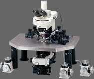
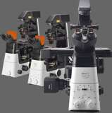
Intracellular recording techniques, such as voltage clamp, current clamp, and patch clamp, require microscopic observation and micromanipulation. These techniques involve penetrating the cell with a very fine glass micropipette (less than one micron in diameter). The delicate micromanipulation of the cell, and the need, in many circumstances to penetrate thick tissues (in order to locate a specific nerve cell, for example), demands a very stable, vibration free microscope configuration with protection from electrical interference. High N.A. long working distance objectives such as water dipping / water immersion objectives are essential for deep imaging and in enabling the use of micromanipulation equipment and electrodes. Useful imaging modes include, brightfield, darkfield, confocal, DIC, IR-DIC, and fluorescence. The use of living cells may require a heated stage or tissue / organ bath on the microscope and other forms of environmental control.
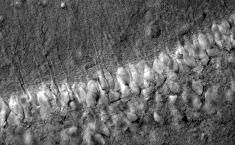
* Image Credit: Dr.Hiroyoshi Miyakawa, Dr.Shigeo Watanabe, Laboratory of Cellular Neurobiology,Tokyo University of Parmacy and Life Sciences, School of Life Sciences.