Nikon Instruments Inc. | Americas
- en Change Region
- Global Site
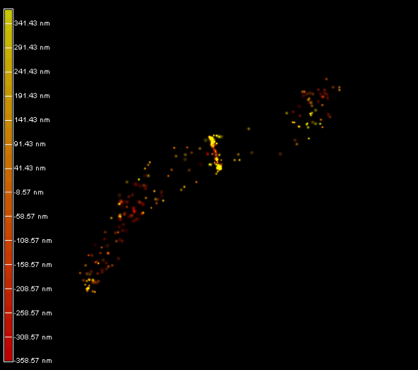
3D-PALM imaging of the FtsZ protein coupled with mEOS2 in Vibrio cholera; Courtesy of Dr. Romain Le Bars (Galli, E. et al. Cell division licensing in the multi-chromosomal Vibrio cholerae bacterium. Nat Microbiol 1, 16094 (2016))
Microbiology is the study of micro-organisms. These include bacteria, yeasts, simple fungi, algae, protozoans, and viruses. Micro-organisms can infect living tissues and may be responsible for causing disease. Being able to identify the infecting agent is the basis for effective treatment. Important techniques in detecting the presence of micro-organisms in cells and tissues (either directly or, most commonly, following in vitro culture) include molecular techniques, immunoassays, and light microscopy. Direct observation of viruses is beyond the resolving power of the light microscope. The presence of viruses in cells can be detected by consequent morphological changes, by using highly specific fluorescently labelled antibodies to specific viral antigens, or by FISH.
Bacteria are among the smallest of light microscope-visible organisms and generally require the use of oil immersion or water-dipping lenses to improve resolution of bacterial shape and cell structure. Contrast can also be improved with the use of non-specific stains (to stain all microbes in the specimen) and specific stains (staining only certain cells or cell structures). The Gram stain is, perhaps, the best know stain and is used to classify bacteria into Gram+ and Gram- bacteria depending on the characteristics of the cell wall (Gram+ have more highly cross linked peptidoglycan with smaller pores enabling retention of dye).
Key clinical microscopy techniques in microbiology include brightfield, darkfield, phase contrast, DIC, oil-immersion, the use of water dipping / water immersion objectives, fluorescence, histology, immunohistochemistry and immunocytochemistry, digital imaging, and telepathology.