Nikon Instruments Inc. | Americas
- en Change Region
- Global Site
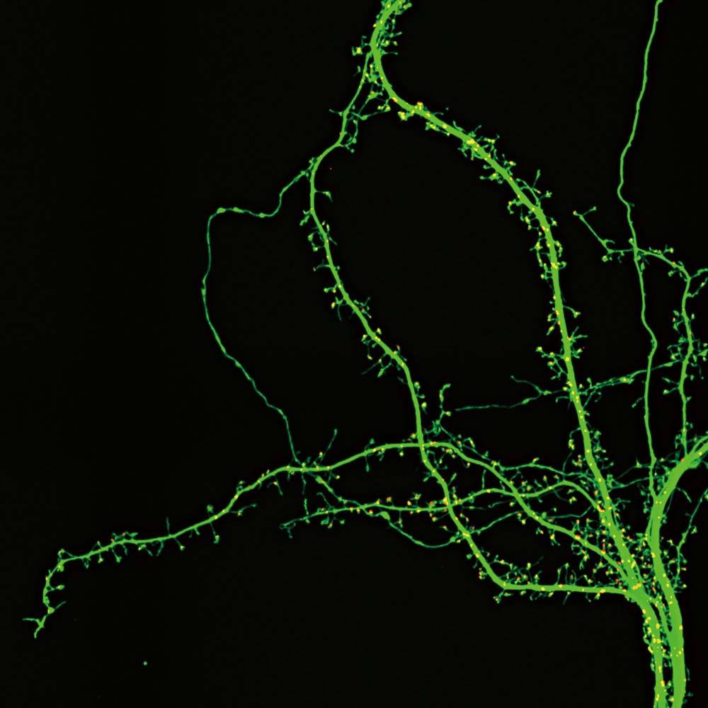
Image courtesy of: Drs. Yutaro Kashiwagi and Shigeo Okabe, Department of Cellular Neurobiology, Graduate School of Medicine and Faculty of Medicine, The University of Tokyo.
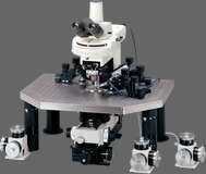
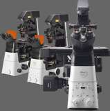
Neurobiology is the study of the cells of the nervous system (principally neurons and glial cells) and the organization of these cells into functional circuits able to process information and mediate behavior. Neuronal circuits can be complex - there are over 100 billion interconnected neurons in the human brain, for example. One of the difficulties in observing neural networks is that single neurons can extend deep into tissues - tissue sections, therefore, provide only limited information. Confocal microscopy, multiphoton imaging, optical sectioning and 3-D reconstruction are powerful techniques that allow cells of the nervous system to be visualised deep into brain slices and other tissues. Similarly, the use of specialized objectives designed for deep imaging, such as water dipping and water immersion objectives, improve results.
The development of brain tissue and neuronal outgrowths can be observed in detail in model organisms with translucent embryos using high-resolution light microscopy (confocal, fluorescence, and DIC) and time-lapse imaging. Gene knock down and enhanced gene expression techniques allow the role of specific neuronal growth factors and other signalling molecules in inducing neuronal outgrowths, synapse formation, and neuronal circuits to be studied. Cell migration can also be studied using fluorescence techniques involving powerful fluorophores, such as photactivatable GFPs, kaede, and quantum dots.
The assembly of microtubules and their interaction with the cell membrane is involved in directing neurotransmitter vesicles to synapses. TIRF microscopy allows direct observations of microtubule function and vesicle movement within approximately 50-100 nm of the plasma membrane, while confocal microscopy can be used to monitor movement in the micron range. The pattern of calcium movement involved in modulating synaptic transmission can be observed with the help of fluorescent / luminescent calcium indicators and techniques such as FRAP and FLIP, while movement of calcium ions across the plasma membrane may be recorded using patch-clamp electrophysiology. The protein-protein interactions leading up to calcium release, in addition, can be monitored using techniques such as FRET and BRET.