Nikon Instruments Inc. | Americas
- en Change Region
- Global Site
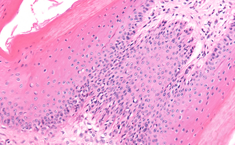
Histology is the study of tissue morphology using light or electron microscopy. Tissue samples are generally obtained through surgery, biopsy or post mortem.
Tissue preparation for light microscopy involves either fixation to prevent decay (using formaldehyde), embedding in wax, and sectioning into 2-8 micron slices with a microtome, or freezing and sectioning with a cryostat. The thin tissue slices are then placed on a glass microscope slide for staining. A wide range of histological stains is available to selectively identify particular cell components. Similarly, fluorescent dyes and immunohistochemical labels can also be used to target specific proteins, carbohydrates, and lipids. FISH may be used to identify specific nucleic acid molecules. Frozen sections are particularly useful when results are required rapidly, for example, during surgery or when target structures are lost in fixation techniques - such as lipids and some antigens.
Key microscopy techniques in histology include brightfield, fluorescence, and immunofluorescence. Ergonomic design is also a key requirement for clinical microscopes. Digital imaging, digital slide, and telepathology technologies greatly assist histologists in archiving slide specimens, storing images, creating an audit trail of specimen examinations, and in obtaining second opinions or returning results to remote locations.
