Nikon Instruments Inc. | Americas
- en Change Region
- Global Site
Haematology is the study of blood, blood-forming organs, and blood disease. Blood is composed of three principal cell types: red blood cells or erthyrocytes (containing haemoglobin and responsible for carrying oxygen to tissues), white blood cells or leukocytes (several different types of cells, for example, granulocytes, lymphocytes, and monocytes with immune functions), and platelets or thrombocytes (involved in blood clotting). These cells are suspended in plasma, which also contains water, nutrients, proteins (importantly clotting factors), hormones, and waste products. Red and white blood cells and platelets are generated mainly in the bone marrow.
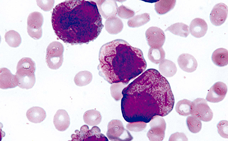
Changes in the structure and number of different blood components can be indicative of blood disorders and other diseases. Many of these changes can be observed in blood and bone marrow preparations (aspirate and biopsy) using light microscopy. Cells are smeared thinly onto glass microscope slides to allow individual cells types to be recognised and counted. A number of stains are available that help in the identification of particular cells and cell organelles. Giemsa, for example, is used in blood and bone marrow preparations and stains red blood cells pink, platelets pale pink, lymphocyte cytoplasm blue, and leucocyte chromatin magenta. Giemsa can also be used to detect blood parasites such as malaria and other spirochete and protozoan microorganisms. Key clinical microscopy techniques include brightfield, darkfield, DIC, fluorescence, immunocytochemistry, immunohistochemistry, digital slide, and telepathology.
