Nikon Instruments Inc. | Americas
- en Change Region
- Global Site
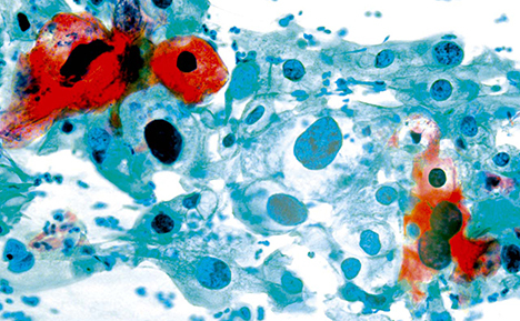
Cytology / cytopathology is the study of cell structure with the aim of diagnosing disease. Cytological screening for disease tends to look for abnormal cells in clinical samples, including, blood, urine, ascites, cell smears (predominantly sources of exfoliated cells), and fine needle aspiration biopsies. Samples are generally prepared by spreading / depositing a thin film of cells onto glass microscope slides. These are then stained (cytological stains, fluorescent stains, and immunofluorescent stains) and examined by a cytologist using light microscopy to identify key cell features, such as cell nuclei, mitochondria, and lysosomes or the presence of micro-organisms such as viruses, bacteria, and fungi. Cytology is most commonly used for the detection of dysplastic cells for cancer screening, particularly for cervical cancer.
Key microscopy techniques in cytology include brightfield, phase contrast, DIC, fluorescence, immunofluorescence, and FISH. Ergonomic design is also a key requirement for clinical microscopes, especially in screening applications. Digital slide and telepathology technologies greatly assist cytologists in archiving specimens, creating an audit trail of observations and in obtaining second opinions, or returning results to remote locations.
