Nikon Instruments Inc. | Americas
- en Change Region
- Global Site

Greater than 70% of investigational new drugs fail during drug development because of unsatisfactory absorption, distribution, metabolism, excretion, and toxicity (ADMET) characteristics. Tools that help to predict the ADMET response early in the development chain are an advantage as they avoid wasting valuable resources on compounds destined to ultimately fail.
Microscopy is increasingly being recognized as a valuable tool in ADMET testing, which can be employed in both in vitro and in vivo preclinical studies. Microscope images can reveal multiple pieces of information (high content imaging) on the cellular response to drug compounds in one experiment. This might include the simultaneous acquisition of data on drug-receptor binding, where this occurs in the cell, and any morphological effects of drug treatment. The ability to use several protein-specific fluorescent probes in one experiment is a key enabling technique in high content imaging.
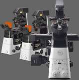
Time-lapse imaging can be used to monitor downstream drug effects and, ultimately, excretion from the cell. Once images have been digitally captured, it is also possible to examine the data retrospectively in response to new questions about the drug compound. Microscopy images have the advantage that they are easily machine readable with appropriate image analysis software. This makes microscopy amenable to medium- to high-throughput analysis (depending on bioinformatics capabilities). Motorized and computer controlled microscopes are essential for automated image capture and for incorporation into medium / high throughput environments.
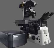
Key techniques include brightfield, darkfield, phase contrast, DIC, fluorescence, confocal, time-lapse, multidimensional imaging, FRET / BRET, FLIM, and FRAP.