


Application Notes
3D Imaging of Kidney Proximal Tubule Microphysiological Systems
December 2023
Microphysiological Systems (MPS) are 3D culture systems that mimic the in vivo environment of live tissues and have been attracting attention in recent years as tools that enable preclinical drug evaluation due to their ability to recreate the physiological environment in the human body. Such human MPS models have shown significant promise in serving as more predictive translational in vitro alternatives compared to using animal models, which are ineffective for predicting clinical outcomes. Nortis‘ ParVivo™ Platform is an MPS that can generate and support tubular tissues, such as blood vessels and renal tubules with continuous perfusion-based fluid flow. By surrounding cells with a biologically relevant matrix, the microenvironment enables research in conditions closer to living tissue than is possible with conventional 2D cultures, where cells are in contact with inorganic surfaces and remain static. Such systems are already used in various fields of research such as regenerative medicine, drug development, toxicity evaluation, and disease research, and are expected to become a new standard in vitro evaluation method. This article demonstrates the observation of primary cilia on human renal proximal tubule cells in a human proximal tubule model constructed with the ParVivo™ Platform using the Nikon AX/AX R confocal microscope. This microscope system captures a wider area with higher resolution 3D structures than conventional confocal microscopes, making it possible to clearly observe small structures such as primary cilia which are approximately 200 nm in diameter.
Keywords: microphysiological system (MPS), organ-on-a-chip, 3D culture, confocal microscopy, proximal tubule, primary cilia, regenerative medicine, drug development, toxicity evaluation, disease research
3D imaging of the proximal tubule model reproduced using the ParVivo™ Platform
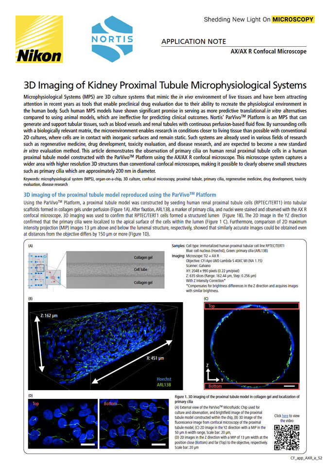
Using the ParVivoTM Platform, a proximal tubule model was constructed by seeding human renal proximal tubule cells (RPTEC/TERT1) into tubular scaffolds formed in collagen gels under perfusion (Figure 1A). After fixation, ARL13B, a marker of primary cilia, and nuclei were stained and observed with the AX R confocal microscope. 3D imaging was used to confirm that RPTEC/TERT1 cells formed a structured lumen (Figure 1B). The 2D image in the YZ direction confirmed that the primary cilia were localized to the apical surface of the cells within the lumen (Figure 1 C). Furthermore, comparison of 2D maximum intensity projection (MIP) images 13 µm above and below the lumenal structure, respectively, showed that similarly accurate images could be obtained even at distances from the objective differs by 150 µm or more (Figure 1D).

Samples
Cell type: Immortalized human proximal tubular cell line RPTEC/TERT1
Blue: cell nucleus (Hoechst), Green: primary cilia (ARL13B)
Imaging
Microscope: Ti2 + AX R
Objective: CFI Apo LWD Lambda S 40XC WI (NA 1.15)
Scanner: Galvano
XY: 2048 x 990 pixels (0.22 µm/pixel)
Z: 635 slices (Range: 162.44 µm, Step: 0.256 µm) With Z Intensity Correction*
*Compensates for brightness differences in the Z direction and acquires images with similar brightness.


Figure 1. 3D imaging of the proximal tubule model in collagen gel and localization of primary cilia
(A) External view of the ParVivoTMMicrofluidic Chip used for culture and observation, and brightfield image of the proximal tubule model constructed within the chip, (B) 3D image of the fluorescence image from confocal microscopy of the proximal tubule model, (C) 2D image in the YZ direction with a MIP in the 50 µm X-width range, Scale bar: 20 µm, (D) 2D images in the Z direction with a MIP of 13 µm width at the position close (Bottom) and far (Top) to the objective, respectively. Scale bar: 20 µm
Example of analysis of primary cilia using General Analysis 3
General Analysis 3 (GA3) is a GUI-based image processing and measurement module in NIS-Elements that can be used to generate custom analysis workflows. Using GA3, we analyzed the orientation of primary cilia by extracting the primary cilia area from the bottom MIP image and calculating the angle at which the Feret’s diameter was largest relative to the direction of media perfusion (Fig. 2A, B, and C). As a result, it was quantitatively shown that the primary cilia in the image were oriented along the direction of perfusion (Figure 2D).


Figure 2. Analysis of primary cilia orientation
(A) Image used for analysis, (B) Area of primary cilia extracted using GA3 (magenta), (C) Calculation method of primary cilia orientation, (D) Circular histogram showing the orientation of primary cilia calculated from the image (n = 85)
ParVivo™ Platform
The ParVivo™ Platform is an innovative technology consisting of a Microfluidic Chip, Perfusion Module, and Perfusion System, that enables reproduction of living tissues mimicking a true physiological environment. The system is characterized by its ability to easily introduce and culture cells in a 3D matrix, generating various tissue models of blood vessels, renal proximal tubules, or the blood-brain barrier within the Microfluidic Chip. In addition, the Perfusion Module and Perfusion System support the long-term preservation of cell survival and function while more accurately reproducing the flow rates and shear forces within living tissues by controlling the amount of perfusion to match the specific tissue at a constant level. In addition, these systems are designed to facilitate microscopic imaging, allowing easy observation of the Perfusion Module during the incubation period or of the Microfluidic Chip following fixation at the end of an experiment (Figure 3).


Step 1. An extracellular matrix such as collagen is injected into the chamber, surrounding the glass fiber. Cells can also be mixed in the matrix.
Step 2: After gel formation, the glass fiber is pulled out to form a tubular perfusion channel in the gel, into which epithelial or endothelial cells are seeded and cultured.
Step 3. Cells attach to the gel to reproduce tubular tissues such as blood vessels and proximal tubules. Drugs and other substances can be added to the tubular tissue from within the lumen or from the extracellular matrix side.
Figure 3. Overview of ParVivo™ Platform
Summary
The proximal tubule model constructed by Nortis’ ParVivoTM Platform was observed using Nikon’s AX/AX R confocal microscope. As a result, the tubular structure of RPTEC/TERT1 formed in collagen gel, as well as the structure and orientation of primary cilia along the direction of perfusion, were clearly captured in all areas of the luminal structure. The ParVivoTMPlatform is capable of reproducingvarious tissues more faithfully to the living body, and Nikon’s AX/AX R confocal microscope offers superior performance for high-resolution, detailed observation and quantitative analysis. Combining these two systems will enable the acquisition of more physiologically-relevant data in the development of disease mechanisms and treatments, and is expected to become a powerful research tool in the fields of medicine, biology, and drug development.
References
S Ishida, T Kanamori, Microphysiological system as a promising technology for drug assay, Folia Pharmacol. Jpn ., 154, 345-351. (2019).
Anna Tourovskaia et al . Brief Communication: Tissue-engineered Microenvironment Systems for Modeling Human Vasculature, Exp. Biol. Med ., 239(9): 1264–1271. (2014). Elijah J. Weber et al . Development of a microphysiological model of human kidney proximal tubule function, Kidney Int ., 90(3): 627–637. (2016).
Devices Used
ParVivo™ Platform (Nortis, Inc.)
The combination of a microfluidic chip, perfusion module, and pneumatic pump supports the creation, culture maintenance, and observation of microphysiological systems. It also enables perfusion of test compounds and live imaging while perfusing.
Product Information
Supports high-speed, high-resolution, wide-field confocal imaging with low phototoxicity to living cells and low light fading.
- High Speed : 720 frames per second
(Resonant 2048 x 16 pixels) - High resolution :
Up to 8K (Galvano) / 2K (Resonant) - High throughput :
Ultra-wide field of view of 25 mm