
Smart Automation with Confocal Microscope: ECLIPSE Ji Changes the Future of 3D Cell Research
AdHuCell platform and Research Institute for Biosciences
University of Mons - UMONS
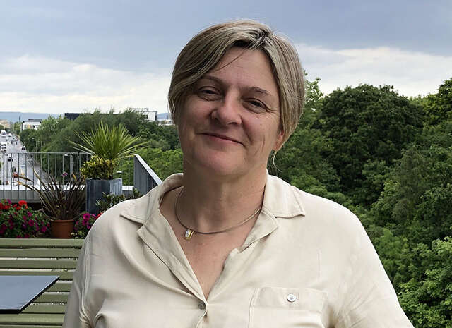
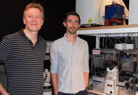
Professor Sylvain Gabriele, SYMBIOSE Lab
Professor Sylvain Gabriele at the University of Mons (UMONS), Belgium, is a leading researcher in mechanobiology, investigating how cells sense, respond to, and retain memory of mechanical forces.
In this interview, we spoke with Professor Gabriel about how the Nikon ECLIPSE Ji-AX Confocal Integrated System has advanced their understanding of complex 3D biological systems.

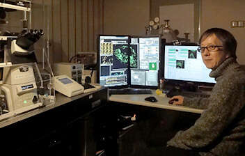
Enduring Excellence - How Long-Lasting Microscopy Fuels Innovation at LIV
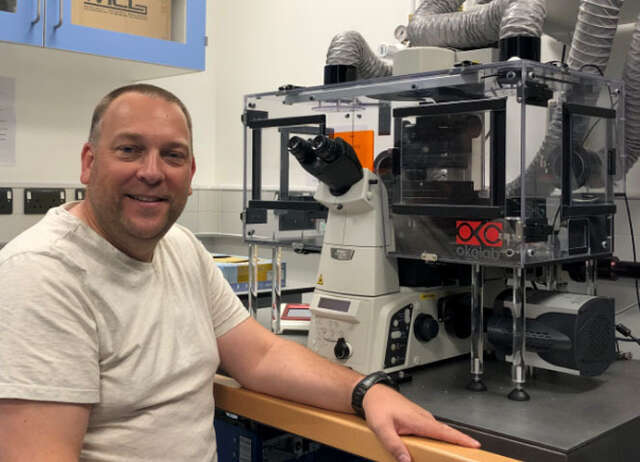
Dr. Christian Conze
Head of Technology Platform Light Microscopy and Image Analysis at Leibniz Institute of Virology (LIV), Hamburg
Celebrating Nikon Microscopy’s 100th Anniversary, our Enduring Excellence campaign honors researchers whose long-term use of Nikon systems showcases the sustainability and lasting value built into our instruments. In this edition, we turn to Christian Conze, Head of the Technology Platform for Light Microscopy and Image Analysis at the Leibniz Institute of Virology (LIV) Nikon Center of Excellence in Hamburg, who has relied on long‑serving Nikon microscopes throughout his research journey.

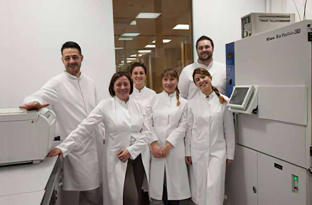
Customer Interview - “This microscope will enable embryologists to perform more ICSI/IMSI procedures per day, and improve embryo quality.”
Dr. David Jareño Martínez
Senior Embryologist, CHIREC IVF Laboratory
Board Member, Belgian Society for Reproductive Medicine (BSRM)
In Belgium — a country known for its highly developed healthcare system within Europe — the CHIREC (Centre Hospitalier Interrégional Edith Cavell) Hospital Group is committed to advancing comprehensive and innovative medical care across a wide range of disciplines.
Belgium is also recognized as the birthplace of ICSI (Intracytoplasmic Sperm Injection), where the world’s first successful procedure was performed.* Building on this spirit of innovation, CHIREC’s IVF (in vitro fertilization) laboratory is dedicated to providing sophisticated and personalized fertility treatments tailored to each patient.
Now, Nikon’s motorized inverted microscope ECLIPSE Ti2-I, designed for ICSI/IMSI procedures, is poised to bring new possibilities in this clinical setting.
We spoke with Dr. David Jareño Martínez, Senior Embryologist at CHIREC’s IVF laboratory for over 16 years, about his impressions of the microscope’s optical performance, automation functions, and the potential impact these may have on treatment outcomes.
* The world’s first ICSI procedure was successfully performed at UZ Brussel.
https://brusselsmorning.com/thirty-years-ago-the-very-first-icsi-baby-was-born-in-uz-brussel/20731/
Disclaimer: This article includes comments from healthcare professionals regarding Nikon products. These comments do not constitute a guarantee of the products’ efficacy, performance, or results.
Furthermore, they do not imply that the healthcare professionals endorse, recommend, supervise, or preferentially select Nikon products.

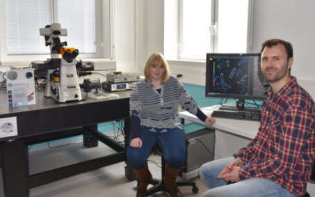
Enduring Excellence - Long-Term Performance in Practice at VIB-KU Leuven
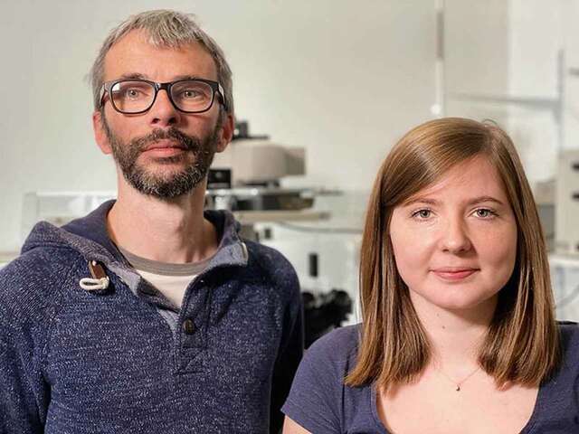
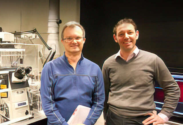
Nikky Corthout
Light Microscopy Expert - VIB BioImaging Core Leuven, VIB-KU Leuven Center for Brain and Disease
Pablo Hernández Varas
Head of Core - VIB BioImaging Core Leuven, VIB-KU Leuven Center for Brain and Disease
For more than a century, Nikon microscope systems have been built to last, and the stories behind their longevity continue to inspire us. In this edition of our Enduring Excellence series, we talk with Pablo Hernández Varas and Nikky Corthout, from the VIB BioImaging Core Leuven at Nikon Center of Excellence at KU Leuven, who have been using the Nikon A1 R confocal microscope system that’s been with the lab for over 17 years. Their insights highlight not just the system’s durability, but how it continues to support cutting‑edge scientific discovery to this day.

“Automating adjustments has reduced wrist and arm strain, making long practice sessions much more comfortable.”
The ECLIPSE Ti2-I supports embryologists training at South Korea’s ART Academy
Eun-Kyung Kim, Ph.D
CHA University Medical Center
Global CHA Embryolab Academy
Fertility Training Center Director
Declining birth rates are a major global issue, and the need for training of embryologists — specialists who handle oocytes, sperm, and fertilized embryos — has become increasingly urgent to help address this problem. The CHA Biomedical Group in South Korea has established the Global CHA Embryolab Academy (GCEA), a specialized institution that systematically trains embryologists to support assisted reproductive technology (ART).
At GCEA, Nikon’s ECLIPSE Ti2-I motorized inverted microscope for ICSI and IMSI plays a vital role for embryologists. Dr. Eun-Kyung Kim, a highly experienced embryologist and director of the Fertility Training Center at the institute, shared insights on the microscope’s operation and the benefits of its implementation.
*This article contains interviews with healthcare professionals regarding our products. However, these interviews do not guarantee the efficacy, effectiveness, or performance of the products, nor do they constitute an official endorsement, recommendation, advice, or selection by the featured professionals.

Sample preparation utilizing the SMZ18 is the key to microscopic videography
YONE PRODUCTION CO., LTD.
Kanako Morioka (Director, Research Department)
Preparing high-quality samples is the first step towards capturing beautiful images. Making samples for microscopy is a detailed, sometimes hours-long process that requires patience, skill, and ingenuity. Yone Production, a company that produces life science videos, employs the SMZ18 stereo microscope for this purpose. Here, Kanako Morioka of the Research Department at Yone Production, who oversees sample preparation, shares her experience of using the SMZ18.

“Automated settings enhance the efficiency of ART in clinical practice, research, and development.”
The ECLIPSE Ti2-I: Contributing to assisted reproductive technology (ART)
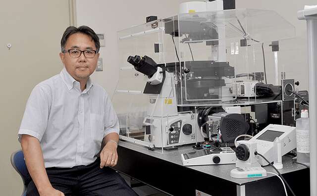
Tatsuya Kobayashi, Ph.D.
Fujita Health University
Fujita Medical Innovation Center Tokyo
The Center for Advanced Reproductive Medicine
Department of Regulatory Science
Associate Professor
The declining birth rate presents a critical social issue that demands attention. Advancements in assisted reproductive technology (ART) are further anticipated to play a key role in addressing this issue. Fujita Medical Innovation Center Tokyo (FMiC) is committed to clinical practice, and the ongoing research and development of ART to help more people wishing to become parents. Nikon’s motorized inverted microscope for ICSI/IMSI, the ECLIPSE Ti2-I, plays a key role in this process. To gain deeper insights into its implementations and practical applications, we spoke with Dr. Tatsuya Kobayashi, an embryologist and Associate Professor at Fujita Health University’s Department of Regulatory Science.
*This page contains interviews with healthcare professionals regarding our products. However, these interviews do not guarantee the efficacy, effectiveness, or performance of the products, nor do they constitute an official endorsement, recommendation, advice, or selection by the featured professionals.

YOUR SWITCH TO THE FUTURE
Department of Biomedicine, University of Basel
Pascal Lorentz
Dr. Michael Abanto
Director of the Nikon Center of Excellence
Head of the Microscopy Core Facility
Research Outline: Immunology and infectious diseases, neurosciences, cancer biology, and tissue development and regeneration.

Dr. Kevin Richetin
Our Nikon Microscopy Ambassador Dr. Kevin Richetin is a neuroscience researcher studying Alzheimer’s disease and the development of tools for neurodegenerative diseases. In this interview video, Dr. Richetin discusses how they chose the Nikon microscope because its software NIS-Elements allows seamless integration of analysis and automation. He emphasizes that this unique capability is incredibly valuable for tracking particles with extremely high resolution over extended periods, a feature that sets Nikon apart.

Customer Interview - A step up in digital pathology using innovative digital microscopy.
Kitasato University Kitasato Institute Hospital
Director, Department of Pathology
Dr. Ichiro Maeda
Currently there are various treatment methods under development that could provide optimal medical care in response to the pathological attributes of each patient. Therefore, the role required of pathological diagnosis, which is indispensable for the planning of treatment policies and strategies, is becoming more important than ever before. However, the shortage of trained pathologists who assume this role is a common issue, not only in Japan, but also worldwide. To solve this problem, digital pathology has been attracting attention. In this article, we asked Dr. Ichiro Maeda, Director of the Department of Pathology, Kitasato University Kitasato Institute Hospital, for his impressions of using the digital image display optical microscope "ECLIPSE Ui".
*Dr. Maeda has provided feedback to Nikon on this microscope’s features and clinical usefulness based on his own personal experience.

"In vitro fertilization is a race against time. I thought this microscope was very helpful in that sense."
In April 2022, insurance coverage for infertility treatment started in Japan, making it easier for couples who want to conceive to receive ICSI treatment. However, in general, there is still a belief that “infertility is not a disease”, so there are many patients who suffer from anxiety or feel impatient during treatment. We interviewed Shoko Ieda and Sumi Shimamura of the Minatomirai Yume Clinic, who say that they find it rewarding to acknowledge these emotions and help such patients with the birth of their babies, about their impressions of using “ECLIPSE Ti2-I”.
*Customers have provided feedback to Nikon on this microscope’s features and clinical usefulness, based on their own personal experience

YOUR SWITCH TO THE FUTURE
Cincinnati Children’s Hospital Medical Center
Matthew Kofron, Ph.D.
Professor, UC Department of Pediatrics
Alicia Ostmann
Cincinnati Cystic Fibrosis Theratyping Research Center
Kentaro Iwasawa, M.D.
Takebe Lab, Division of Gastroenterology
Research Outline: Child health improvement. Organoid creation. Drug efficacy for cystic fibrosis patients.

YOUR SWITCH TO THE FUTURE
European Institute for Molecular Imaging (EIMI)
Professor Friedemann Kiefer, Ph.D.
Department of Intravital Molecular Imaging
Research Outline: Elucidation of how molecular mechanisms shape human cells and tissues biologically. Multiscale imaging.

YOUR SWITCH TO THE FUTURE
National Institutes of Natural Sciences, National Institute for Physiological Sciences
Tetsuhisa Otani, Ph.D. Assistant Professor
Division of Cell Structure
Research Outline: Epithelial tissue.

YOUR SWITCH TO THE FUTURE
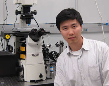
Korea Advanced Institute of Science and Technology
Professor Won Do Heo, Ph.D.
Department of Biological Sciences
Research Outline: Optogenetic and bio-imaging technologies.

YOUR SWITCH TO THE FUTURE
Chinese Institute for Brain Research, Beijing, China
Dr. Hu Zhao, Principal Investigator
Research Outline: New tissue clearing methods, and building a neural connectome mapping platform based on these methods.

Kundeninterview - „Ich glaube, dass dies die herkömmliche Mikroskopie für Pathologen verändern kann“
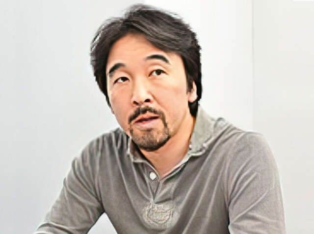
Takeshi Sasaki
Professor, Projektleiter, MD Ph.D
Universitätskrankenhaus Tokio
Abteilung für Pathologie, Informationsvernetzung der nächsten Generation
In der Medizin spielen Pathologen eine äußerst wichtige Rolle bei der Bestätigung von Untersuchungen. Sie schauen oft über lange Zeiträume hinweg in ein Mikroskop, was eine große körperliche und geistige Belastung darstellt. Um diesen Stress für Pathologen zu reduzieren, hat Nikon ein neues Mikroskop für pathologische Untersuchungen entwickelt, das ECLIPSE Ui. Mit diesem neuen Mikroskop können Pathologen hochaufgelöste Bilder von Proben auf einem Monitor betrachten, anstatt durch Okulare zu schauen. Hier berichtet Dr. Takeshi Sasaki vom Tokyo University-Hospital, der dieses Mikroskop getestet hat, über seine Eindrücke.
*Basierend auf seinen eigenen persönlichen Erfahrungen hat Dr. Sasaki Nikon Feedback zu den Funktionen und der Nützlichkeit für klinische Anwendungen dieses Mikroskops gegeben.

Nikon BioImaging Lab contributes to “mini-gut” research
Dr. Hidenori Akutsu
Director of the Department of Reproductive Medicine, Center for Regenerative Medicine, National Center for Child Health and Development
Nikon Equipment and Services

YOUR SWITCH TO THE FUTURE
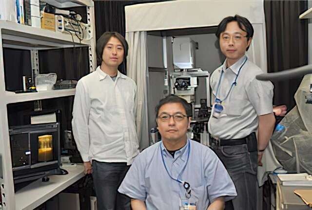
Research Institute for Microbial Diseases (RIMD) at Osaka University
Hiroaki Miki, Professor
Yosuke Funato, Associate Professor
Osamu Hashizume, Assistant Professor
Division of Cellular and Molecular Biology, Department of Cellular Regulation
Research Outline: Analyzing the function of a membrane protein molecule called Cyclin M, which plays a role in ejecting magnesium ions from cells.

Mit dem ECLIPSE Ei im Kurssaal die Ausbildung unterstützen
“Für mich ist es einfach, den Schülern beizubringen, wie man es benutzt, und genauso einfach ist es für die Schüler, es zu benutzen.”
Beim Studium der Grundlagen der Naturwissenschaften spielt die Mikroskopie eine äußerst wichtige Rolle. Das Nikon ECLIPSE Ei wurde als Ausbildungsmikroskop mit dem Konzept entwickelt, ein “benutzerfreundliches Mikroskop zu sein, gerade für Studenten, die zum ersten Mal ein Mikroskop bedienen sollen“. Wir besuchten Frau Rika Izumi, Dozentin für Naturwissenschaften an der Rikkyo Niiza Junior and Senior High School, Saitama, Japan, wo das ECLIPSE Ei in den Kursräumen verfügbar ist. Von ihr wollten wir mehr erfahren über Hintergründe und Begründung für die Wahl dieses Mikroskops, die Erfahrungen der Benutzer sowie die Reaktionen der Studentinnen und Studenten.

The ECLIPSE Ci-L plus contributes to more comfortable clinical examinations.
„Mir wurde sofort klar, dass die hohe optische Leistung im Einklang mit dem durchdachten Design unsere tägliche Arbeit unterstützt.“
Klinische Untersuchungen spielen eine wichtige Rolle in der medizinischen Versorgung. Ein klarer Fokus bei der Entwicklung des neuen Labormikroskops ECLIPSE Ci-L plus von Nikon lag auf dem Konzept, sowohl die physische als auch mentale Belastung von Klinikern und Labortechnikern, die täglich viel am Mikroskop arbeiten, zu reduzieren. In diesem Interview sprachen wir mit Dr. Akira Yoshikawa von der Abteilung für anatomische Pathologie des Kameda Medical Center – einem führenden Lehrkrankenhaus im südlichen Teil der Präfektur Chiba, Japan – über seine Gedanken nach dem Arbeiten mit diesem Mikroskop und den neuen Objektiven „CFI Plan Apochromat Lambda D“ im praktischen Alltagsgebrauch.
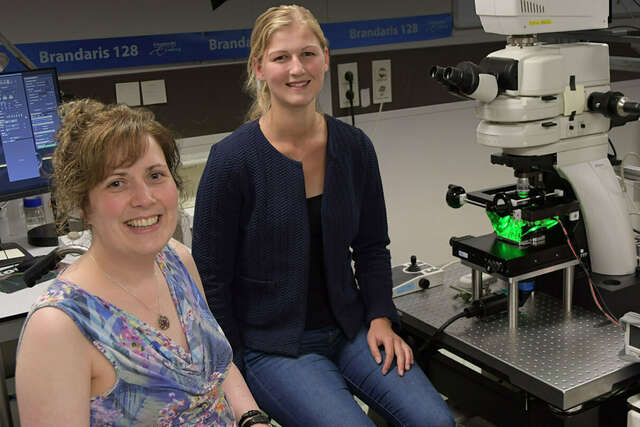
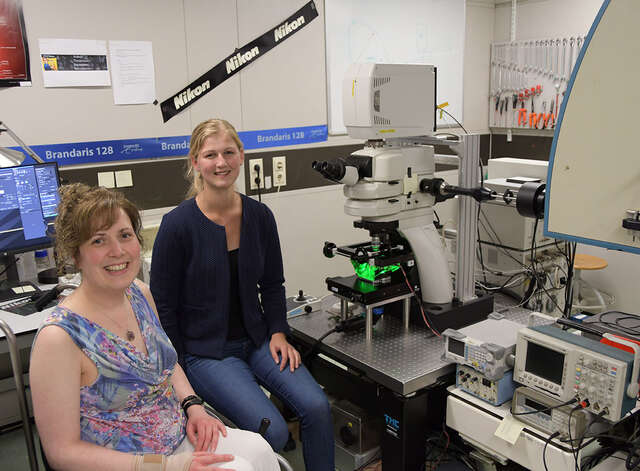
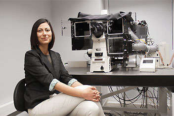
Associate Prof. Dr. Klazina Kooiman and Dr. Ines Beekers
Associate Prof. Dr. Klazina Kooiman, Head of Therapeutic Ultrasound Contrast Agent Group, and Dr. Ines Beekers, Postdoctoral Researcher in the Department of Biomedical Engineering of the Thoraxcenter, Erasmus MC, Rotterdam, the Netherlands

Reproduktionsklinik Tokio
„Das beste Sperma unter Millionen finden. Für Embryologen ist dies eine einzigartige Arbeit der Selektion und wir fühlen eine große Verantwortung dabei.“
Die Reproduktionsklinik Tokio, eine Kinderwunschklinik im dritten Stockwerk des Shiodome City Center, einem hochmodernen Bürogebäude im Shiodome-Viertel im Zentrum von Tokio, hat viele Patienten, die auch an Wochentagend abends auf eine Behandlung warten. Wir sprachen mit Shimpei Mizuta und Tomohiro Maekawa, die das Inverse Mikroskop Ti2 für die Spermiensortierung und die intrazytoplasmatische Spermieninjektion (ICSI) verwenden, über ihre Gedanken zu ihrer Arbeit und ihre Eindrücke vom Ti2.
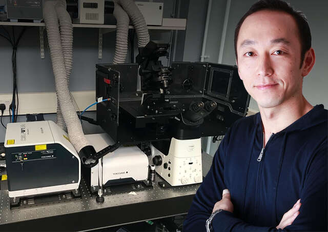
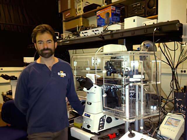
Dr. Yohei Yamauchi
Dr. Yohei Yamauchi, Principal Investigator, Cell biologist of viral infections, School of Cellular and Molecular Medicine, University of Bristol, UK
Assistant Prof. Joseph Michael Hyser & Dr. Alexandra Leigh Chang-Graham
Virology and Microbiology
Baylor College of Medicine
Houston, Texas, USA
Assistant Prof. Klazina Kooiman & Inés Beekers
Therapeutic Ultrasound Contrast Agent Group, Thoraxcenter, Department of Biomedical Engineering, Erasmus MC, Rotterdam
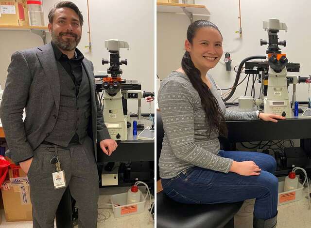
Dr. Steven Nedellec and Dr. Tiphaine Douanne
Dr. Steven Nedellec, Facility Manager of MicroPICell, Université de Nantes, France and Dr. Tiphaine Douanne, Universite de Nantes, Signaling in Oncogenesis, Angiogenesis and Permeability, CRCINA INSERM U1232, France
Dr. Steve Thomas
Senior Lecturer in Cardiovascular Science, University of Birmingham
Dr. Tadahiro Iimura
Division of Bio-Imaging, Proteo-Science Center (PROS), Ehime University
Division of Analytical Bio-Medicine, Advanced Research Support Center (ADRES), Ehime University
Graduate School of Medicine, Ehime University
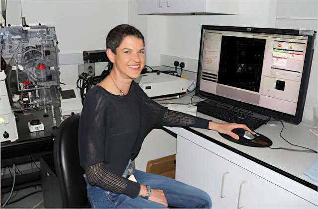
Melike Lakadamyali, Ph.D.
The advanced Fluorescence Imaging and Biophysics Group, ICFO-Institute of Photonic Sciences
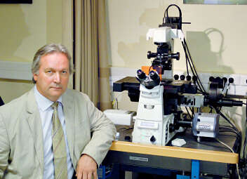
Simon C. Watkins, Ph.D.
Professor and Vice Chairman of the Dept. of Cell Biology
Director and Founder of the Center for Biologic Imaging
University of Pittsburgh
Pittsburgh, Pennsylvania, USA
Ronald D. Vale, Ph.D.
Professor and Vice-Chairman of the Department of Cellular and Molecular Pharmacology
Investigator, Howard Hughes Medical Institute (HHMI)
The University of California, San Francisco San Francisco, CA, USA
Tomomi Nemoto, Ph.D. and Ryosuke Kawakami, Ph.D.
Research Institute for Electronic Science
Sapporo, Hokkaido, Japan
Tamas Freund, Ph.D. and Istvan Katona, Ph.D.
Institute of Experimental Medicine of the Hungarian Academy of Sciences (IEM HAS)
Budapest, Hungary
Maddy Parsons, Ph.D.
Group Leader (Royal Society University Research Fellow)
Randall Division of Cell and Molecular Biophysics
King’s College London
London, United Kingdom
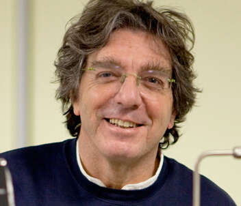
Alberto Diaspro, Ph.D.
Professor of Applied Physics, Department of Physics, University of Genoa
Director of the Department of Nanophysics, Istituto Italiano di Tecnologia
Paul Ronald Selvin, Ph.D.
Professor, Department of Physics, University of Illinois at Urbana-Champaign
Bo Huang, Ph.D.
Assistant Professor, Department of Pharmaceutical Chemistry, Department of Biochemistry and Biophysics, University of California, San Francisco
Atsushi Miyawaki, M.D., Ph.D.
Senior Team Leader, Laboratory for Cell Function Dynamics, RIKEN Brain Science Institute
Ikuo Wada, Ph.D.
Department of Cell Science, Institute of Biomedical Science, Fukushima Medical University
Romain Le Bars, Ph.D.
The Imagerie-Gif light microscopy core facility is a member of the France Bioimaging Infrastructure. The facility is hosted by the Institute for Integrative Biology of the Cell (I2BC) at Gif sur Yvette, France.
Prof. Staffan Strömblad, Ph.D.
Staffan Strömblad Ph.D. is group leader at the prestigious Karolinska Institutet, Stockholm, Sweden, an institution that awards the Physiology Nobel Prize yearly. He is also the head of the Live Cell Imaging Facility (LCI), in which the Nikon Center of Excellence for live cell imaging is integrate.
Dr. Arne Seitz, Dr. Romain Guiet and Thierry Laroche
The Faculty of Life Science (SV) at the École Polytechnique Fédéral de Laussane (EPFL), Switzerland, has a long record of excellence in research applied to life sciences
Dr. Jacopo Lucci
Chief Scientific Officer at Natural Bio-Medicine SpA,
Aboca Group
Masato Nakagawa
So-called iPS cells are attracting considerable interest as pluripotent stem cells that may open up a whole new world of medicine. The Center for iPS Cell Research and Application (CiRA) at Kyoto University is pursuing a wide range of research activities that aim to realize regenerative medicine utilizing iPS cells. The Nikon BioStation CT cell culture observation system is being used in this iPS cell research and is contributing to its efficiency.
We were pleased to have had an opportunity to speak with Masato Nakagawa, who is engaged in iPS cell research at CiRA.
Prof. Heinz Beck, Ph.D.
Full time professor at the Laboratory of Experimental Epileptology and Cognition Research hosted in the Life & Brain Center, Part of the University of Bonn.
Note: The institutions and job titles listed with each researcher reflect their affiliation at the time of the interview.
- Home
- Hintergrundwissen
- Interviews mit Kunden