Ikuo Wada, Ph.D.
Department of Cell Science, Institute of Biomedical Science, Fukushima Medical University
Nikon-Ausrüstung
- N-SIM Super-Resolution
- A1Rsi Confocal (see current model)

What kind of research are you engaged in?
I have long been interested in the mechanism by which secreted proteins are efficiently produced. To study this, I have conducted a series of studies into the dynamics of inner membrane proteins and the dynamics of protein molecules themselves.
Initially, I used confocal microscopes only for observation of immunostained samples, but since starting live cell imaging, I use various techniques such as FCS and TIRF. I want to observe the cells in their living state, and I have a boundless interest in the dynamics of cell structure and molecules.
Why were you interested in the N-SIM super resolution microscope?
Initially, I was anticipating that I would be able to clearly observe the microstructures of endomembranes such as endoplasmic reticulum that cannot be resolved well with a confocal microscope even with high magnifications. When I used it in practice, I discovered that this was not the only thing I could now do-and when I used it in combination with an A1 confocal microscope system, it felt as if a whole new world had been opened up.
How did you find the N-SIM to use in practice?
I’m not a physics specialist, so I read about SIM (structured illumination microscopy) with difficulty and was expecting it to be difficult to use the Nikon N-SIM. But once I started using it, I found it to be extremely user-friendly and resolution beyond the diffraction limit can be obtained with simple button operation. It is simple to modify the analysis parameters, and I admire the fact that it allows an intuitive understanding of the correlation between these parameters and the results of data acquisition. It is so reliable that we can reconstruct accurate images while changing parameters after acquisition, as all the original acquisition data are preserved with Nikon’s imaging system.
I was also particularly struck by the N-SIM’s superb resolution in the Z-axis direction compared with that of confocal microscopes.
How have you been using the A1 and the N-SIM in combination?
I mainly use the N-SIM for observing cell structures as still images. On the other hand, the A1 can perform ultrahigh-speed image acquisition and allows FRAP and photo stimulation. Unless both devices are used, the information we can obtain is limited.
The combination of N-SIM and A1 provides us with a wide range of imaging possibilities. For example, it allows acquisition of both wide-area images in low magnification using A1 and high-resolution images of detailed structures using N-SIM.
What are the advantages of mounting analysis units such as FCCS and FLIM to the A1?
First, the A1 is highly extensible and customization, including external inputs, is extremely simple. This makes it easy to use-even for an amateur of optics like me. We use FLIM mainly for measurement of FRET efficiency during intramolecular structural changes. Accurate measurement of FRET is not easy, and also there are sometimes difficulties with the acceptor-bleaching method, but with FLIM, results are obtained clearly with few artifacts appearing.
I think SIM shows the high performance of Nikon’s optical system, which accurately reproduces theoretical values. It seems that this high optical performance enables us to obtain much higher photon counts per molecule (CPM) than those acquired with other optics with FCS. It is marvelous, because the 60x water-immersion objective that I use tends to produce dark images and lower CPM values. I believe that this image brightness is a great advantage with TCSPC measurement.
What developments would you like to see for N-SIM in the future?
I hope to see the development of simultaneous acquisition of two colors. Even dual-view display with a single camera is pretty handy, though it has only 256 pixels in the horizontal.
I’d also like to see an easier-to-understand explanation of the SIM image reconstruction processes.
Please describe your impressions of Nikon.
I highly appreciate the fact that Nikon accepted my requests on improving the system. Instead of responding to my difficult requests by saying it can’t be done, Nikon is seriously considering how to bring them to fruition. I’m also pleased that Nikon products are outstanding in terms of system extensibility.
The March 11 earthquake struck Japan soon after delivery of this system to our lab. The CO2 incubator fell off the table, but the system wasn’t damaged and could be used after adjustment of settings. I believe the robustness of the system is very important for users.
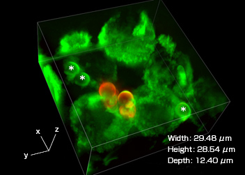
Three-dimensional image of phagosomes taken with a super resolution microscope
J774/mVenus-SNAP-23 cells observed using a super resolution microscope (Nikon’s N-Structured Illumination Microscopy System). Phagosomes (highlighted with green fluorescence), which incorporate beads wrapped in mVenus-SNAP-23 (which are marked with a *), are visible in the cells. After Phagocytosis, the addition of an Alxa594-labeled antibody that recognizes the IgG used in opsonization allows confirmation that there are beads outside the cells (highlighted with red fluorescence).
System:
N-SIM + A1Rsi + FLIM and FCCS unit (PQ device attached to the A1)