


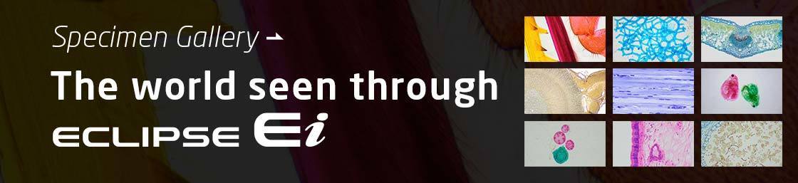
Curiosity – the genuine desire to study the world and unlock its mysteries – is the starting point of science and technology.
The ECLIPSE Ei educational microscope offers unique digital and design solutions to ensure the smooth progress of your courses. Using the ECLIPSE Ei to foster your students’ curiosity and maintain their enthusiasm for learning, you can unlock their potential and open their eyes to the world around them.
Students can enjoy learning how to operate the Eclipse Ei using its smartphone-friendly Online Guide. Digital cameras that enable sharing of discoveries and experiences through images are also available to stimulate your students’ intellectual curiosity.
By simply scanning a QR code, students can quickly access the Online Guide on their smartphones to independently learn how to operate the microscope. The Online Guide can also be useful in preparing students before using the microscope and to reinforce concepts after using the microscope. To access the Online Guide directly, click here.
Read the QR code using a smartphone
Check the Online Guide
Operate the microscope
You can select the Quick Guide, which offers video lectures on basic operations, as well as a Contents List that introduces details about operations and the cleaning procedure.
The Digital Sight 1000 optional microscope camera is simple to operate and ideal for use with the Eclipse Ei. Not only can users record images and videos of specimens, but they can also simultaneously observe images with others via a monitor or network.
By using a monitor to display an image under observation with the Eclipse Ei, a group of people can observe the same specimen simultaneously.
Digital Sight 1000 camera can be easily connected to a large display for classroom lectures and seminars.
By connecting Digital Sight 1000 to a PC equipped with the free software package NIS-Elements LE, you can share images observed with the ECLIPSE Ei in real time with remote PCs or smart devices.
Additionally, by using common web conferencing services, it can serve as an effective tool for online learning.
Images under observation with a microscope can be shared in real time, with students even in distant locations.
(Figure above shows utilization of a web-conferencing service.)
Both ease of understanding and operability, which respectively enable intuitive and stress-free operation, are in demand. Our unique design expertise has been applied to every aspect of the microscope. The accompanying Online Guide empowers students to learn how to operate the microscope independently, revolutionizing the conventional class workflow and saving time for practical training.
The body of the Eclipse Ei is designed to provide smooth and seamless operation which facilitates quick observation. Simple and intuitive markings on the Eclipse Ei, from illustrations to color-coding, enable students to quickly understand how to operate the various aspects of the microscope.
The power switch and light intensity control knob are all located at the front.
You can check which objective is in use and change magnifications without other objectives getting in the way.
You can adjust the eye-point height for a comfortable observation posture by moving the eyepieces up or down.
The handles for moving the stage in the X/Y direction are displayed using illustrations of their respective shapes.
The simple stage shape has no bothersome projections on either side.
The coarse/fine focus knobs for moving the stage up and down are positioned on both sides of the microscope.
The lever positions of the condenser aperture are color-coded and matching the objectives.
Stage height limits can be set to prevent specimen and objective collision and damage.
The tube can be rotated to reduce the space required for storage by loosening the tube locking screw. The tube is designed so that it does not fall.
The Eclipse Ei features a compact, light-weight body that saves space and is easy to handle, as well as a robust design that is built to last. It is easy to carry and store, reducing the stress of setting it up and storing it.
* Compared to previous halogen lamp models
The compact footprint saves valuable space at the bench or desk. In addition, the tube can be rotated towards the back to minimize storage space.
The AC adaptor can be stored at the back of the microscope. The power cord can also be wound up when storing.
Its dramatically reduced weight and multiple grip locations make the Eclipse Ei easy and stable to lift, carry and store, even on high shelves.
A security wire slot is provided and supports maintenance.
Nikon has been developing and refining optical technology since its founding in 1917. The ECLIPSE Ei features Nikon’s high quality optics resulting from over 100 years of optical expertise.
The dedicated CFI BE2 Plan Achromat series objective and the 10X eyepiece achieve a large field of view of 20*, enabling students to find their target specimen or structure more quickly. Infinity-corrected objectives with excellent image flatness and chromatic aberration correction provide high-contrast images that accurately capture the color and shape of specimens.
* 120% or more than that of previous models.
The bright, high-resolution 100X objective* exhibits superior imaging performance through oil immersion, clearly capturing fine structures. The 60X objective* does not require oil immersion for high magnification observation.
* Optional
100X oil immersion objective
60X objective
The illumination system is equipped with a fly-eye lens to achieve uniform brightness across the entire field of view. Low heat generating, long-life LEDs are utilized as the light source.
Fly-eye lens
The user can select either a binocular set or a trinocular set. The trinocular set comprises a camera port with a built-in 0.55X zoom lens. Both sets feature two-stage eyepoint height adjustment.
Eclipse Ei binocular set
Eclipse Ei trinocular set
A color camera equipped with a 2-megapixel CMOS image sensor that can acquire images of up to 1920 x 1080 pixels. Simply connecting it to a monitor* and a mouse enables you to capture images without the need for a PC. In addition to still images, it also enables acquisition of movies and simple measurements such as length and angle on a monitor. By connecting the camera to a PC, you can share specimen images being observed with the microscope with other PCs and smart devices. This makes the Digital Sight 1000 perfect not only for recording images, but also for online education and discussions.
* Via a HDMI cable.
The Digital Sight 1000 can be used with just a monitor and mouse.
Simple measurements such as measuring the distance between two points can be performed on the monitor.
* Calibration by an optional objective micrometer is required.


