Nikon Instruments Inc. | Americas
- en Change Region
- Global Site
The Nikon FX format CMOS image sensor enables instantaneous capture of images in high definition. Digital Sight 10 allows the unprecedented high resolution of 6K and switching color and monochrome capture with a single camera. This high-performance model also features a high frame rate for fast focusing on high-definition images.
Note: All images on this page are examples from research applications. The Digital Sight 10 is not registered for clinical use.
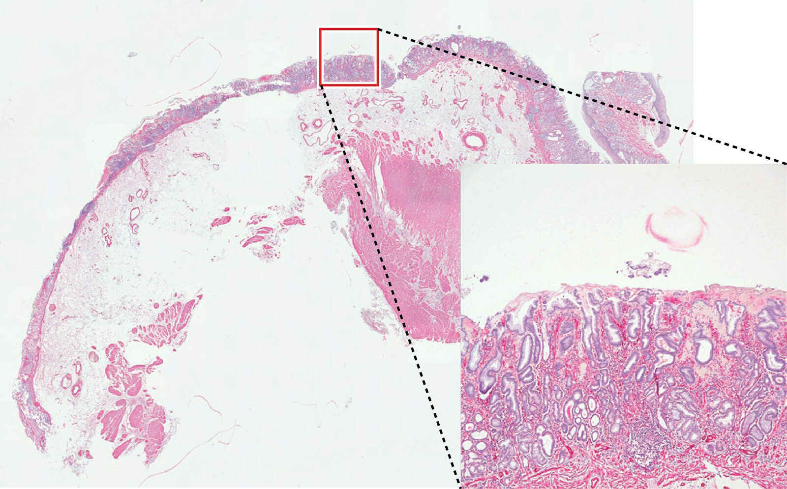
Stomach, SMA staining, 17x12 Tiled images (Objective: CFI Plan Apochromat 40XC)
Photo courtesy of : Nichirei Biosciences Inc.
A 25 mm field of view (FOV), possible in combination with inverted microscopes, and upright microscopes, enabling the capture of images over a wider area in one shot. Tiled images can be created efficiently, cutting the time required for screening.
*Upright microscopes are supported only by the ECLIPSE Ni Series (brightfield).
Digital Sight 10
Conventional model (DS-Ri2)
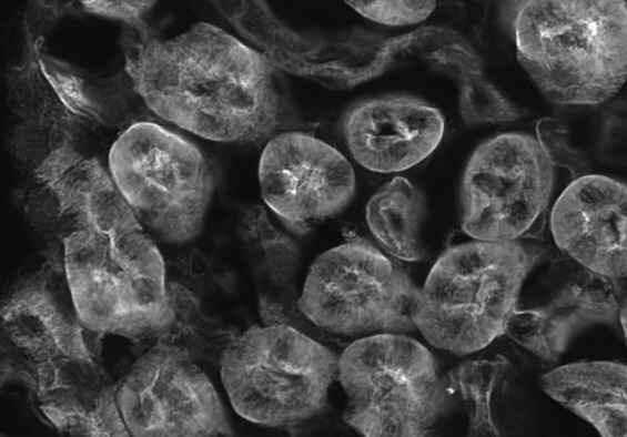
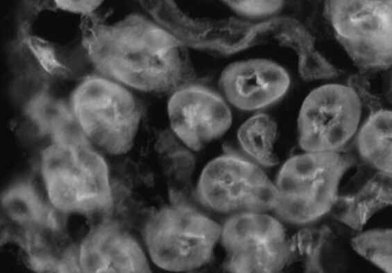
Kidney tissue (WGA: 488) (Objective: CFI Plan Apochromat VC 20X)
Microscopic images can be captured at up to 6000 x 3984 pixels (23.9 megapixels), ideal for image analysis and observation of fine structures.
Digital Sight 10 is capable of live display of 6000 x 3984 pixel (23.9-megapixel) images at 9 frames/second or 1920 x 1080 pixel (2.1-megapixel) images at 66 frames/second. Fine focusing is easy and stress-free. By using the ROI mode, it is possible to shoot only any place at a higher speed.
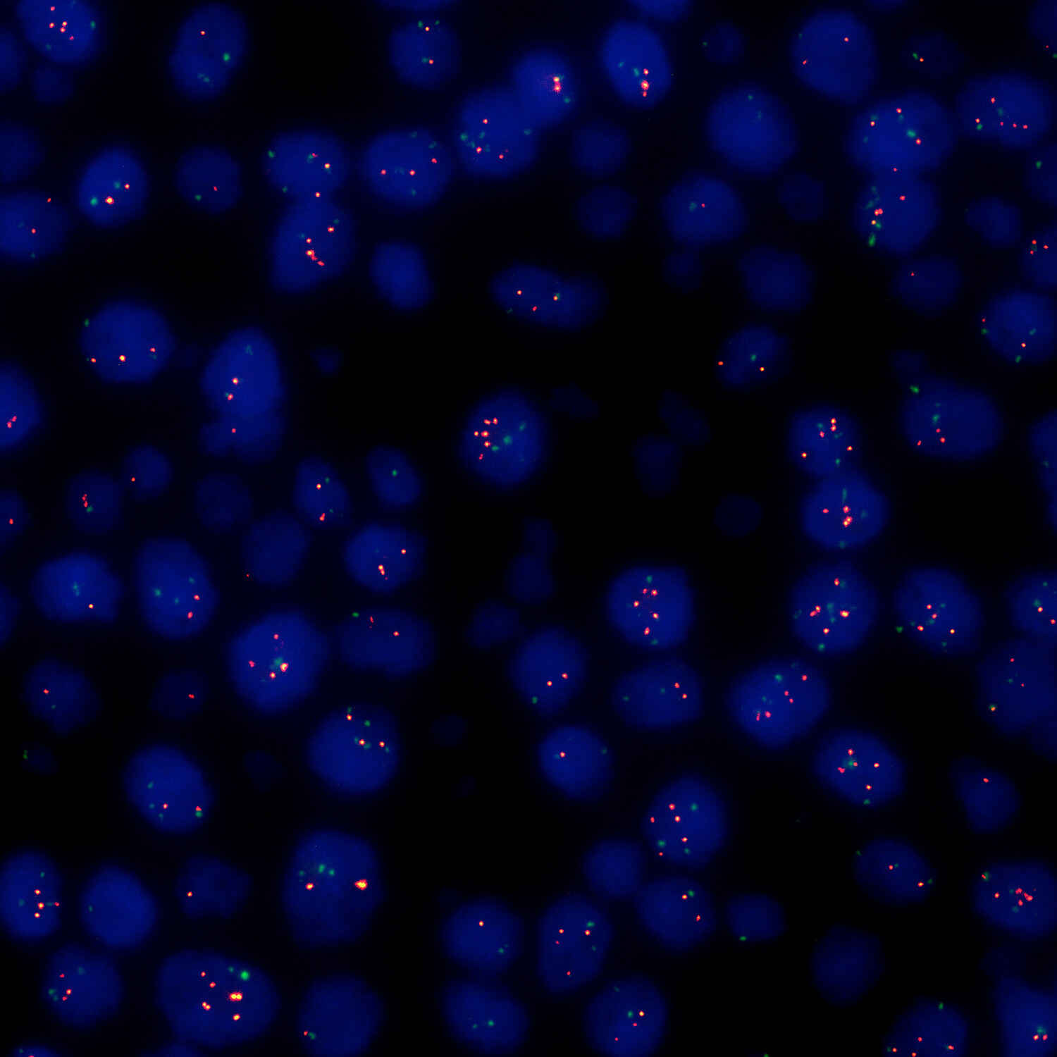
Breast cancer, FISH method (Objective: CFI Plan Apochromat Lambda D 100X Oil)
Photo courtesy of: St. Marianna University Hospital
Digital Sight 10 achieves high sensitivity equivalent to ISO 200 in color mode and ISO 800 in monochrome mode. Clear fluorescence observation with a high signal-to-noise ratio is possible in both monochrome and color image acquisition.
Color Mode
When inserting the color filter can shoot 400 to 680 nm in color
Monochrome mode
When detaching the color filter capable of shooting 400 to 850 nm in Monochrome
*Replace with monochrome IR filter
Easy color mode switching, either manually or electronically
Digital Sight 10 makes it possible to easily switch the color mode either electronically or manually by using specialized imaging software for electronic switching or attaching/detaching filters to the slot at the bottom of the microscope camera for manual switching.
1x electronic adapter
Switch with a single action in the imaging software
*A 1x electronic adapter and a separate PC equipped with specialized imaging software, NIS-Elements, are required for electronic operation.
A single sensor captures both color and monochrome images, for consistent appearance even when switching color mode. Easy image acquisition is possible without the hassle of using different cameras.
Brightfield (monochrome)
Brightfield (color)
Fluorescence (monochrome)
Fluorescence (monochrome)
Zebrafish (Objective: SHR Plan Apo 1X)
Digital Sight 10's monochrome mode supports near-infrared (700 nm) fluorescence image capture, normally difficult to achieve with conventional color cameras. As fluorescence sensitivity extends to the NIR region, this camera is suited to fluorescence image capture of thick samples and samples with weak phototoxicity.
Upright microscope system ECLIPSE Ni
Objective lens for biological microscope Lambda D
Kidney cancer, Vimentin staining (Objective: CFI Plan Apochromat Lambda D 20X)
Photo courtesy of: Nichirei Biosciences Inc.
Blurring and color bleeding are low even to the periphery, for images that are clear even when enlarged. ECLIPSE Ni supports everyday observation and inspection of samples with high resolution and high color fidelity.
Inverted microscope system ECLIPSE Ti2
Objective lens for biological microscope Lambda D
Mouse neuron (Objective: CFI Plan Apochromat Lambda D 40XC)
From captured images of 18 μm thickness every 0.2 μm. Image processed with Clarify.ai
ECLIPSE Ti2 takes advantage of a wide field of view (field number 25) to achieve high throughput even when capturing 3D or other large-size data. Combined with image processing, it enables the capture of clear images with a higher signal-to-noise ratio, even deep into subjects.
Stereoscopic microscope system SMZ25/18
Zebrafish larva (brightfield/myocardium GFP) (Objective: SHR Plan Apo 2X)
Photo courtesy of: Dr. Hiroyuki Nakajima, National Cerebral and Cardiovascular Center
SMZ25/18 offers high definition at high frame rates. Capture perfect, bright images without missing high-speed biological reactions. Low noise makes this system ideal for time lapse imaging.
Nikon uses the NIS-Elements series as control software. NIS-Elements allows functions from basic imaging to control of the microscope and peripheral devices to be performed, as well as the measurement, analysis, and management of acquired images. Four basic packages and a variety of optional modules are available to suit every application and objective.
The bundled free package offers functions for the display of scale on live images, full-screen display, and more. The simple operation screen makes shooting easy.
The documentation package is equipped with measurement and report creation functions. It enables general microscopic image acquisition in fields from biomedical to industrial, and is expandable through optional added features such as EDF and databases.
The research package enables the construction of advanced image acquisition systems, including multidimensional imaging (up to 4 dimensions for Br, 6 dimensions for Ar), through integration with systemized microscopes. Sets equipped with a rich range of image processing and analysis functions are available for every application.
Compatible OS: Windows® 10 and 11 64-bit Professional
* For information about compatible desktop PCs, contact Nikon.