Products and Promotions may differ based on your selected Region.
Return to the top of the page
Discontinued Products
Nikon BioImaging Labs provide contract research services for microscope-based imaging and analysis to the biotech, pharma, and larger research communities. Each lab's full-service capabilities include access to cutting-edge microscopy instrumentation and software, but also the services of expert biologists and microscopists, who are available to provide quality cell culture, sample preparation, data acquisition, and data analysis services.
Search and filter over 125,000 Open Access Articles that utilize Nikon products and supported third party systems.
Input appropriate parameters to calculate important performance specifications such as resolution, depth of field, sampling rate, confocality, and more.
Download software and firmware programs for Nikon microscope products.
Step-by-step guides for aligning and operating select Nikon microscopes.
Nikon's MicroscopyU is a top source for educational information about optical microscopy.
Free interactive microscopy e-Learning courses, including product-specific courses.
Find the right Nikon objective to fit your workflow.
A comprehensive resource for understanding modern microscopy techniques and technologies.
Glial cell surrounded by axons in a rat neuronal culture labeled for microtubules and actinDr. Christophe Leterrier, NeuroCyto, INP, Marseille, France
Pascal LorentzDr. Michael Abanto
Director of the Nikon Center of ExcellenceHead of the Microscopy Core Facility
Research Outline: Immunology and infectious diseases, neurosciences, cancer biology, and tissue development and regeneration.
Matthew Kofron, Ph.D.Professor, UC Department of Pediatrics
Alicia Ostmann Cincinnati Cystic Fibrosis Theratyping Research Center
Kentaro Iwasawa, M.D. Takebe Lab, Division of Gastroenterology
Research Outline: Child health improvement. Organoid creation. Drug efficacy for cystic fibrosis patients.
Professor Friedemann Kiefer, Ph.D.
Department of Intravital Molecular Imaging
Research Outline: Elucidation of how molecular mechanisms shape human cells and tissues biologically. Multiscale imaging.
Dr. Hu Zhao, Principal Investigator
Research Outline: New tissue clearing methods, and building a neural connectome mapping platform based on these methods.
Professor Won Do Heo, Ph.D.
Department of Biological Sciences
Research Outline: Optogenetic and bio-imaging technologies.
Tetsuhisa Otani, Ph.D. Assistant Professor
Division of Cell Structure
Research Outline: Epithelial tissue.
Hiroaki Miki, ProfessorYosuke Funato, Associate ProfessorOsamu Hashizume, Assistant Professor
Division of Cellular and Molecular Biology, Department of Cellular Regulation
Research Outline: Analyzing the function of a membrane protein molecule called Cyclin M, which plays a role in ejecting magnesium ions from cells.
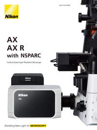
The origin of NSPARC is in the IIT (Italian Institute of Technology). We interviewed Professor Alberto Diaspro, Dr. Paolo Bianchini, and Dr. Giuseppe Vicidomini, who worked on research and development at the Nikon Imaging Center and the Molecular Microscopy and Spectroscopy Lab in the IIT.
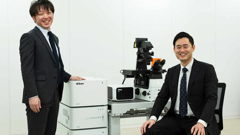
NSPARC enables super-high-speed scanning with unprecedented high resolution for observation and acquisition of images, showing even the slightest of changes in living cells. Its developers spoke of their ambitious challenge of aiming higher.
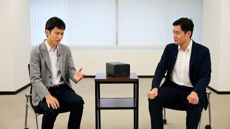
The AX/AX R confocal microscope system is designed to let anyone customize freely according to their usage. To find out more, we spoke to those in charge of product planning and design about the stories behind how they pursued software operability, as well as hardware performance improvements.
Note: The institutions and job titles listed with each researcher reflect their affiliation at the time of the interview.
Download 16.85MB