- en Change Region
- Global Site
- Home
- Products
- Upright Microscopes
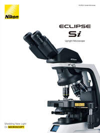
- ECLIPSE Si
Interviews
Customer Interview Contributing to the future of ophthalmic care — the ECLIPSE Si
“I chose it because it’s compact in size, easy to operate and provides us with beautiful images.”
The Nikon biological microscope ECLIPSE Si is designed based on ergonomics, pursuing operational efficiency while maintaining natural posture. Ishizuchi Eye Clinic in Niihama City, Ehime Prefecture, Japan has introduced 'direct microscopy examination of eye discharge', a technique which utilizes the ECLIPSE Si for diagnoses of eye diseases such as conjunctivitis. We interviewed Dr. Takashi Suzuki, the director of the Ishizuchi Eye Clinic, about his reasons for choosing this microscope and its usability.
*This page features an interview with a doctor about a Nikon product. This interview does not guarantee the efficacy, effectiveness or performance of the product, nor does it indicate that the doctor endorses, recommends to instruct with, or select the product.
Contents
Nikon’s ECLIPSE Si supports ophthalmic medical care
The ECLIPSE Si microscope installed in the clinic
The Ishizuchi Eye Clinic, located in Niihama City, Ehime Prefecture, Japan is engaged in ophthalmological medical care rooted in the community. The clinic is operated by a team of accomplished staff members, including several expert ophthalmologists such as the director Dr. Takashi Suzuki, as well as certified orthoptists who specialize in eye examination and training to improve patients’ eyesight. Introducing cutting-edge scientific equipment and surgical tools, the facility provides advanced, wide-ranging medical care that expands the possibilities for patient care choices.
Dr. Takashi Suzuki
Director of the Ishizuchi Eye Clinic, Associate Professor of the Eye Center at the Omori Medical Center of Toho University, Advanced Treatment for Eye Disease class and Endowed class
* Job title and responsibilities are as of the time of interview
One representative example of Dr. Suzuki's activities is his introduction of direct microscopy examination of eye discharge. This test is extremely effective for diagnosing conjunctivitis, a type of eye infection, and contributes to faster, more accurate treatment. The essential equipment for direct microscopy examination of eye discharge is a biological microscope, which is used to check whether bacteria or viruses are in the eye discharge. Dr. Suzuki is currently employing the Nikon ECLIPSE Si for this purpose.
About the direct microscopy examination of eye discharge using the ECLIPSE Si
Direct microscopy examination of eye discharge is mainly used for diagnosing conjunctivitis. Conjunctivitis can have bacterial, viral, or allergic origins, and the treatment methods and prescription drugs vary for each. A normal medical examination might reveal inflammation of the conjunctiva*1, but not necessarily the true underlying cause. If this underlying cause is not identified immediately and accurately, treatment can be delayed and symptoms prolonged.
For the examination, the patient's eye discharge is collected with a cotton swab or similar, applied to a slide glass, then stained to highlight specific cells and organisms during observation under a microscope. By identifying bacteria, inflammatory cells, etc., the cause of the condition can be clearly identified. Through this examination, an ophthalmologist can accurately explain the situation and prescribe appropriate treatments to assist the patient towards a speedy recovery.
Collecting eye discharge (sample)
Observing the stained eye discharge sample with a microscope
Identifying the causative bacteria from the microscope image
We asked Dr. Suzuki about the direct microscopy examination of eye discharge and the role of the ECLIPSE Si.
Low stage for easy sample placement and swapping.
— What kind of equipment is required for direct microscopy examination of eye discharge?
Dr. Suzuki: Basically, it requires a biological microscope with a digital camera attached to it. If you also have a monitor to project the captured images, that would be great. Then, you just need a cotton swab to collect the eye discharge, a slide glass, an alcohol lamp to fix the sample (eye discharge), and a staining kit. You don’t need anything more special than that.
— How long does it take to conduct the test? What skills are required for it?
Dr. Suzuki: Usually, the examination only takes about 10 minutes. How to identify bacteria and inflammatory cells using a microscope can be learned through short-term practical training. Also, this test can have medical fee points attached equivalent to a test such as OCT*2 that requires dedicated equipment. This is a little-known fact.
Confirmation of stage position and observation through the eyepieces is possible while maintaining a comfortable position.
Diagnosis, treatment and prescription based on test results
— What were your reasons for introducing the ECLIPSE Si?
Dr. Suzuki: It was compact in size and didn't interfere with anything else when placed on a desk in the clinic. It seemed easy to operate and provided us with beautiful images. Other reasons are that in actual use, it is easy to set and observe samples, all of which helps us to achieve faster testing.
Nikon’s biological microscope ECLIPSE Si is designed based on ergonomics, so it allows users to maintain a comfortable posture. Since the staging position is low, it is easy to place or change the samples. The position and size of operational parts such as focusing knobs are also designed with ease of use in mind to promote more efficient operation. It also addresses sudden brightness changes that occur when changing the magnification of the objective lens. The Light Intensity Management (LIM) function remembers the illumination brightness setting used for each objective lens, eliminating tedious light adjustments, reducing strain on the eyes caused by glare, and assisting clear imaging. In addition to all these benefits, the ECLIPSE Si is designed with a variety of new ideas to increase functionality and operability.
*1 The conjunctiva is the mucous membrane that covers the back of the eyelids and the surface of the eyeball up to the circumference of the iris.
*2 OCT (Optical Coherence Tomography) is an imaging technique that uses light waves to capture cross-sectional images of the retina.
Contributing to society through improved ophthalmic medical care
Dr. Suzuki explained what he does after conducting the direct microscopy examination of eye discharge.
“I examine the patient's eye at the same place I conduct my medical consultation. While showing images to the patient, I can explain the name of the condition, the treatment method, and the prescription drugs to be given,” he describes. “By delivering faster and more accurate diagnoses, patients can feel safer, and I can gain their trust.”
When an ophthalmologist explains a disease or condition to a patient, the first thing he or she wants to know is often, 'Is it infectious?' or 'How long does it take to heal?' In that situation, being able to positively identify the cause means the doctor can deliver a clear answer.
Microscope images captured by the ECLIPSE Si (example)
Bacteria (circled in red) in eye discharge
Virus-infected cells can be seen in the eye discharge
In order to spread awareness and know-how about this test to ophthalmologists and clinics nationwide, Dr. Suzuki serves as an instructor of skill transfer* sponsored by the Japan Association for Ocular Infection, participates in lectures in various areas and constantly disseminates information on social media.
Dr. Suzuki aims to contribute to the advancement of ophthalmology by facilitating wider recognition, understanding, and adoption of this direct microscopy examination of eye discharge, and helping to support the eye health of as many patients as possible.
When we asked about his future goals, he answered, “I’d like to continue utilizing microscopes to improve our ability to diagnose eye infections.”
Nikon will continue to respond to that passion.
* 'Skill transfer' refers to the handing down of a certain technology or skill to those who do not yet possess it.
Note: The institutions and job titles listed with each researcher reflect their affiliation at the time of the interview.


