The perfect platform for live sample high content.
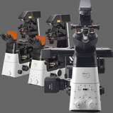
We chose our inverted microscope platform for high content imaging because it was the most flexible: the largest selection of objective lenses, detectors, modalities and applications available to apply to high content acquisition and analysis.
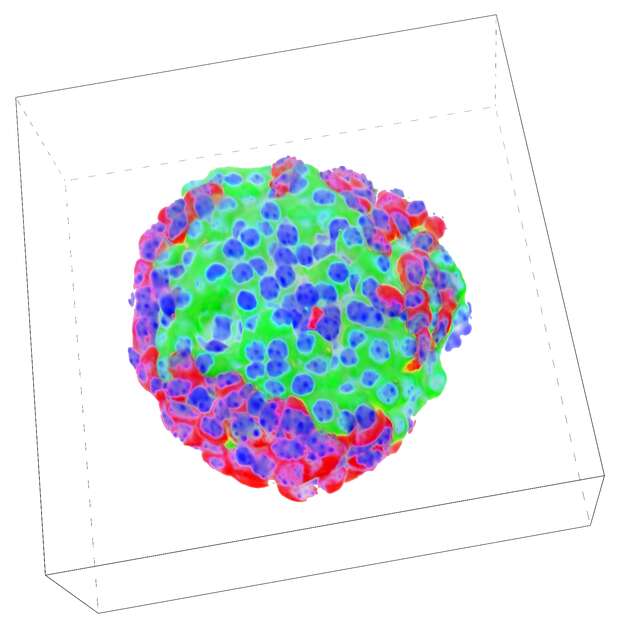
With confocal modalities, 3-dimensional high content at the highest resolution can be acquired. And with space for 44 well plates, multiple users can take advantage of time-resolved live imaging applications.

Key Features
Environmental stability is maintained by a 44-plate capacity incubator (two stackers each housing up to 22 plates) and a full imaging enclosure.
With a main door for plate loading, and a side door for direct robotic transfer to the microscope stage, temperature and humidity can be quickly recovered and constantly maintained.
An internal robot picks plates and loads them through a side door to a second robot which transfers plates directly to the microscope stage, all inside an incubated environmental chamber.
Automated imaging on a schedule

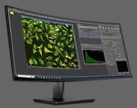
The NIS-Elements Scheduler allows users to maintain their individual plates and schedule open times on the imaging system to run them.
Always running, the scheduler shows an overview of all plates and their owners.
The Scheduler automatically loads plates at imaging time and executes the experiment defined by the user. When complete, the plate is returned to the incubator.
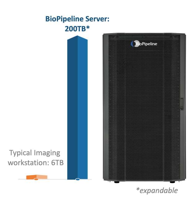
Storage Capacity
High content imaging can mean large storage requirements. Having over 30x the storage capacity of a typical high-end imaging workstation, with expandability for more, the included BioPipeline Server with direct dedicated 10GbE network connection allows for more uninterrupted imaging and offline processing and analysis.
BioPipeline’s data transfer rates from the imaging workstation to the dedicated server exceed most traditional networks by 10-100x by using dedicated 10 GbE connections.
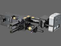
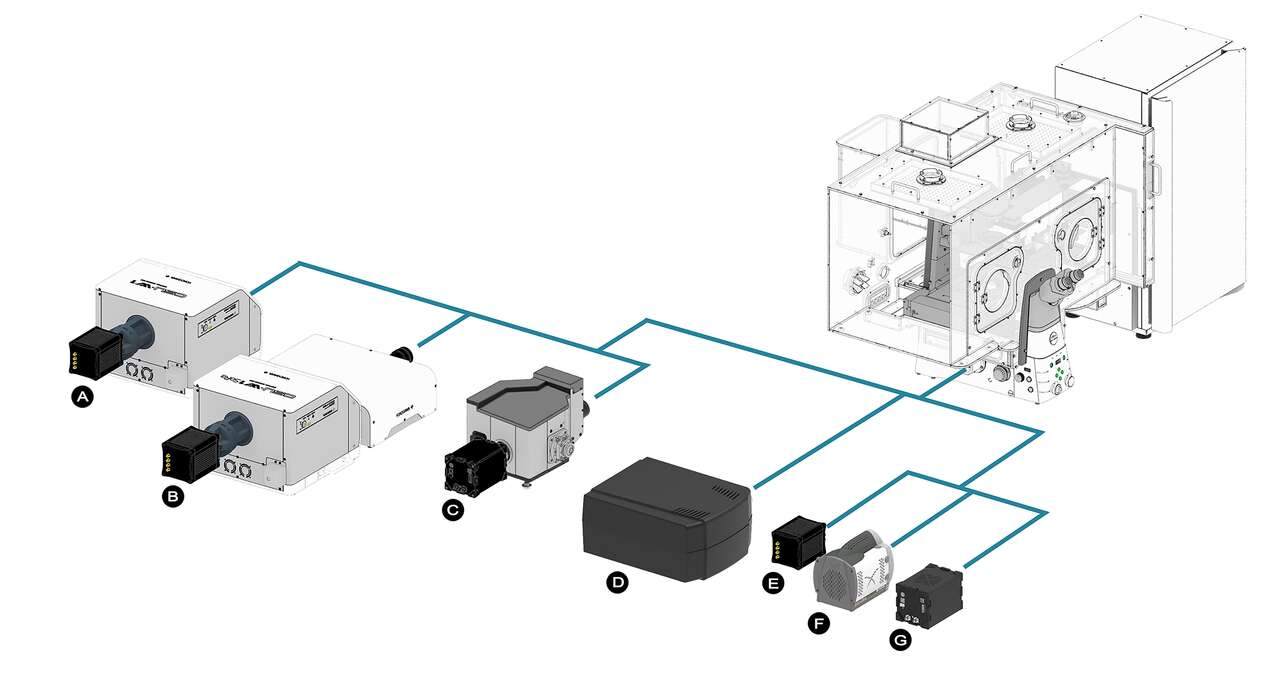
Detector Choice
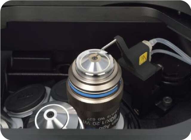
Options for detectors is one of BioPipeline’s most useful features, allowing users to customize the detector type to the assays being performed, and allowing an upgrade path for future detectors.
There are also multiple detector ports that can be utilized on the microscope.
Several options are available:
A. Spinning disk confocal
B. Super-resolution spinning disk confocal
C. Large FOV spinning disk confocal
D. Large FOV point scanning confocal
E. One or more sCMOS cameras
F. One or more EMCCD cameras
G. One or more Large FOV sCMOS cameras
Increase analytical throughput without compromising image resolution
The combination of a high-speed resonant scanner and large field of view forms an ideal platform for high-resolution screening assays. It dramatically reduces the time needed to analyze multiple samples and conditions.
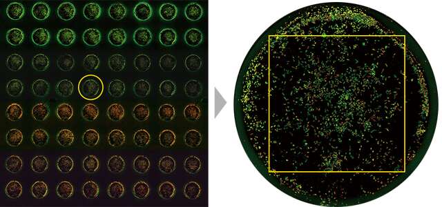
Left: A well in the 96-well plate is selected
Right: The large FOV enables acquisition of entire wells (with a 4X objective)
Large FOV enables measurement of larger areas and high-throughput analysis
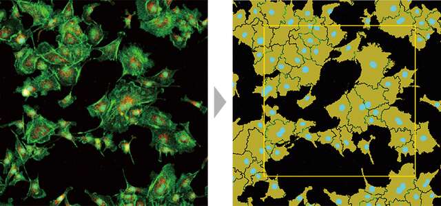
Analysis Toolbox
Image analysis either during or post-acquisition is an essential tool for high content imaging. NIS-Elements includes several analysis tools as well as capabilities for user-defined custom assays that can be applied to images.
Analysis results can be used as well to direct the course of experiments in real-time.
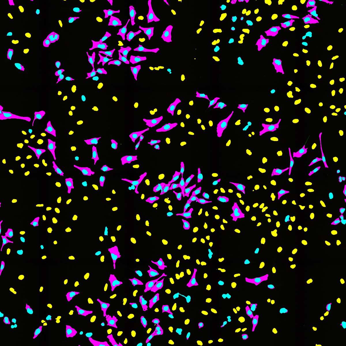
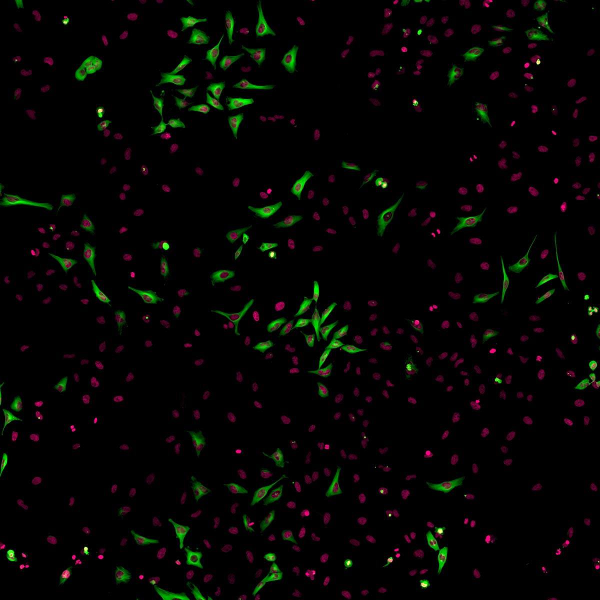
Cell classification tool segments and classifies samples into categories for counting and measurement
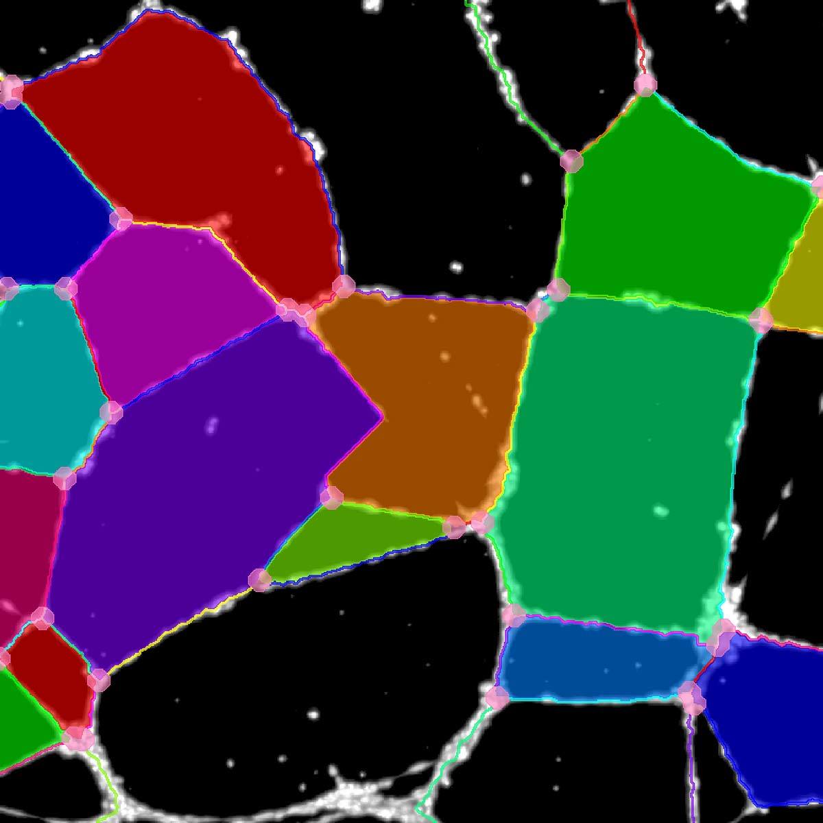
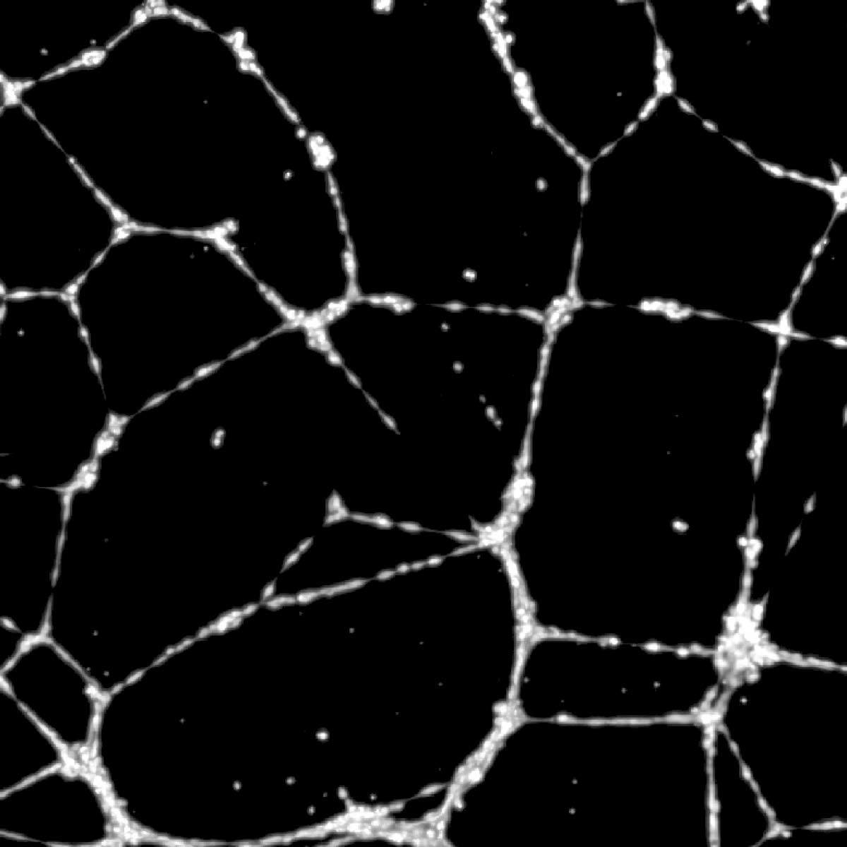
Angiogenesis tool detects tube formation and measures tube number, area, nodes, segment number, and segment lengths.
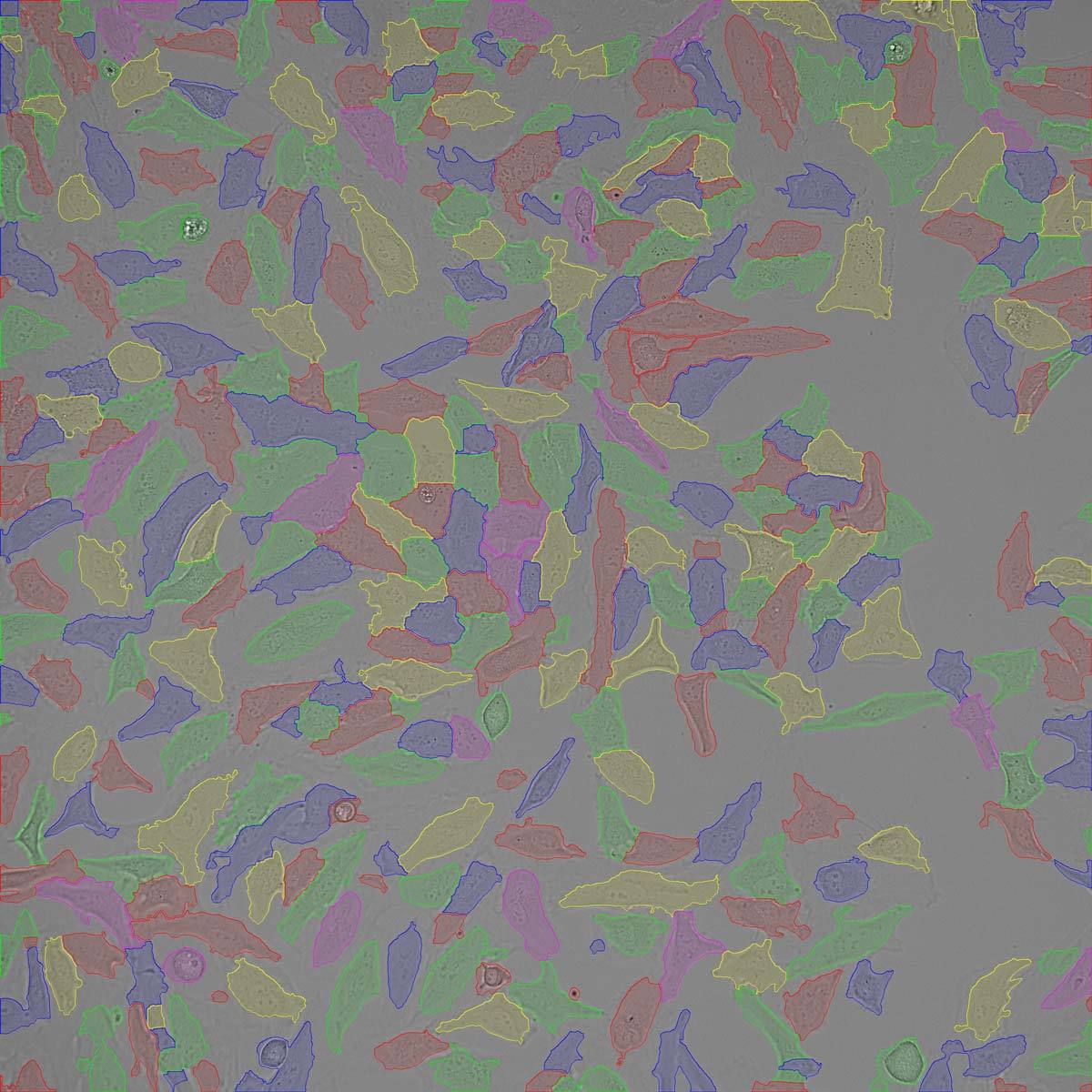
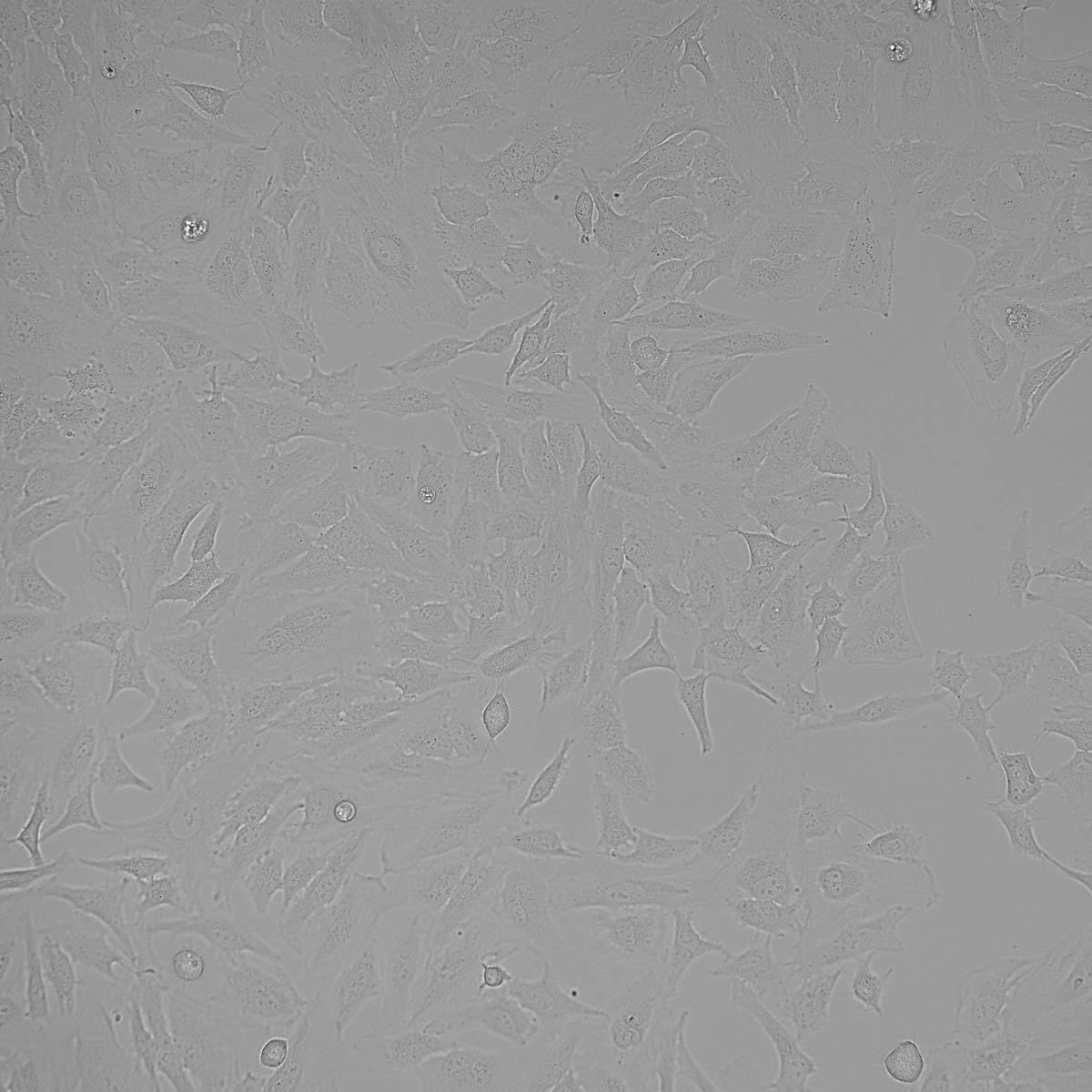
Cell counting tool using standard brightfield imaging to segment and count cells.
Product Brochure
High Content Imaging
- BioPipeline LIVE
- BioPipeline PLATE
- BioPipeline SLIDE
- ECLIPSE Ji