 ®
®
Nikon Instruments Inc. | Americas
- en Change Region
- Global Site

The BioPipeline SLIDE system utilizing the Marzhauser Slide Express 2 loader provides an extremely robust and stable platform for automated slide scanning.
With several detector and objective lens choices, high quality images can be obtained and stored in a fully automated fashion.
* BioPipeline Slide is not for clinical diagnostic use.
By using a cassette with precision slide mounting, slides are loaded with care and repeatability. This means true automated runs of up to 120 slides.
Because of the slide mounting mechanism, flatness across the slide is also maintained, speeding up total acquisition time by negating the need for repeated autofocus over long distances.
Multichannel fluorescence mouse embryo acquired by tiling multiple fields of view acquired with a 20x objective lens
For slides with specimens mounted randomly, a low magnification preview can locate the sample, then acquire only the sample at higher magnification and high resolution. Not acquiring the entire slide saves time and space. Users then have the tiled high magnification images (red square shows one FOV in the tiled image, shown at right)
Low magnification preview (not saved)
High magnification tiled images are acquired
An individual frame of the tiled image
Image analysis either during or post-acquisition is an essential tool for high content imaging. NIS-Elements includes several analysis workflows as well as capabilities for user-defined custom assays that can be applied to images.
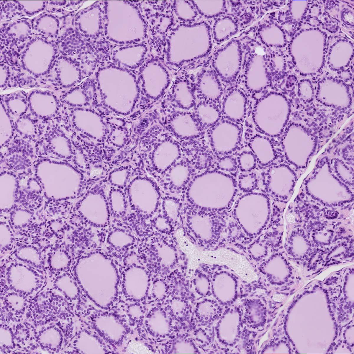
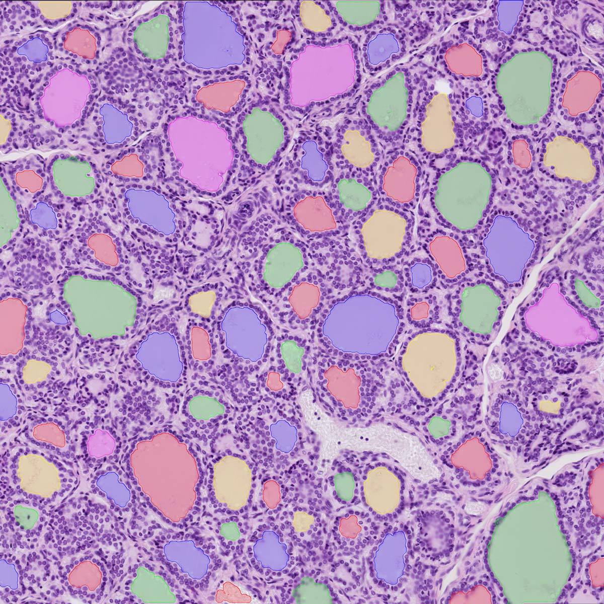
An example of a custom segmentation tool to detect colloids in tissue sections.
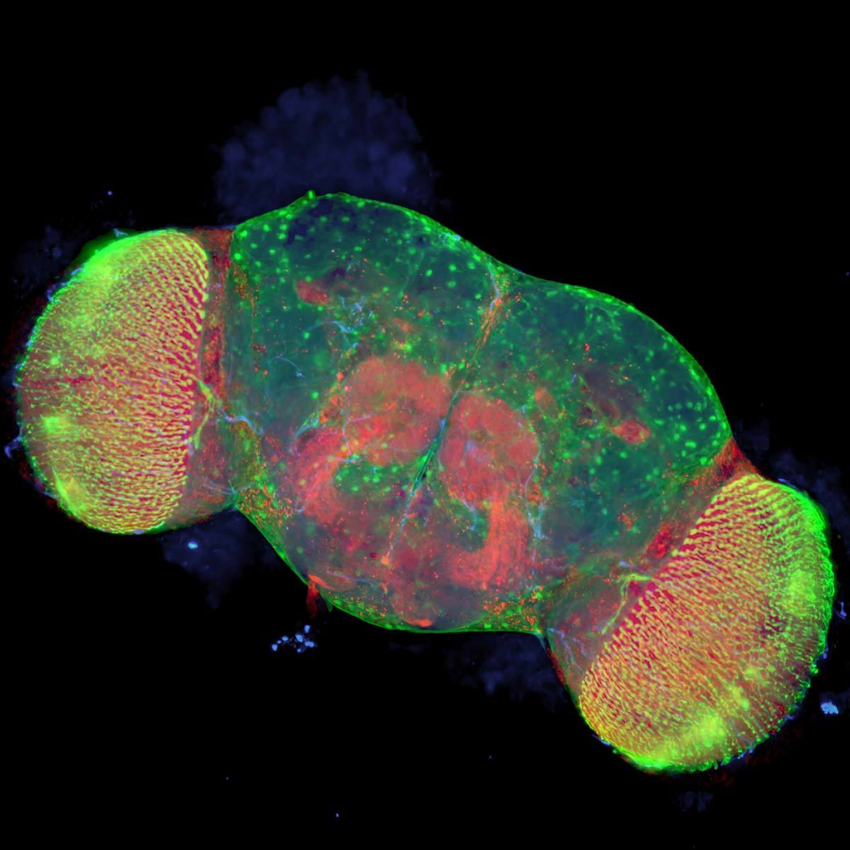
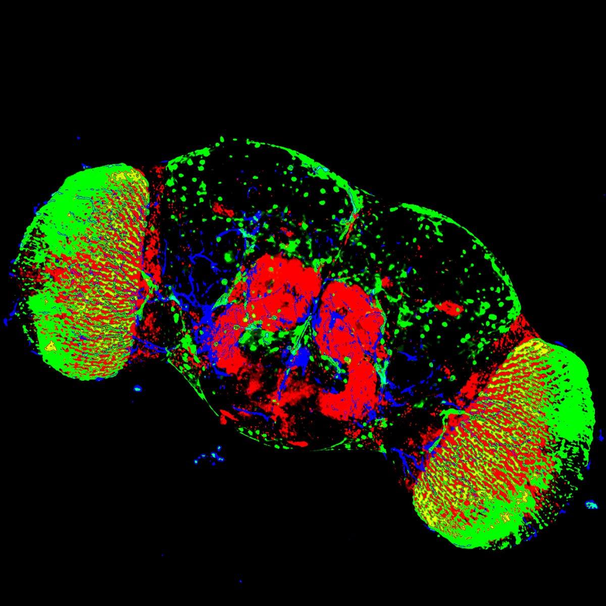
Fluorescence color segmentation tool to determine percent area occupied by each fluorescence channel.
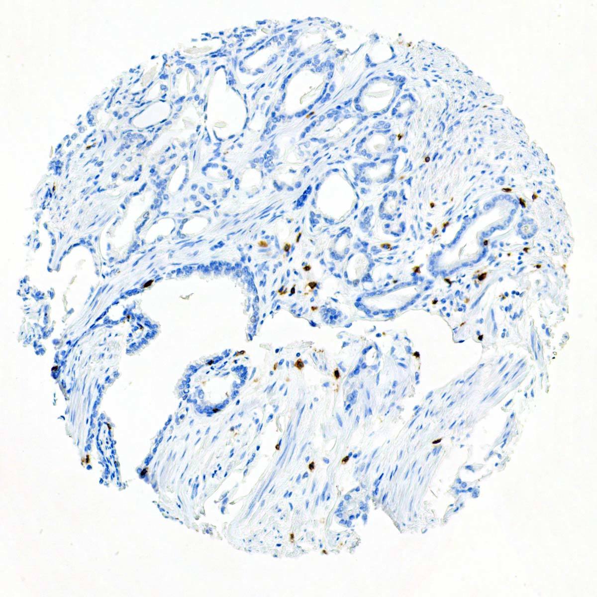
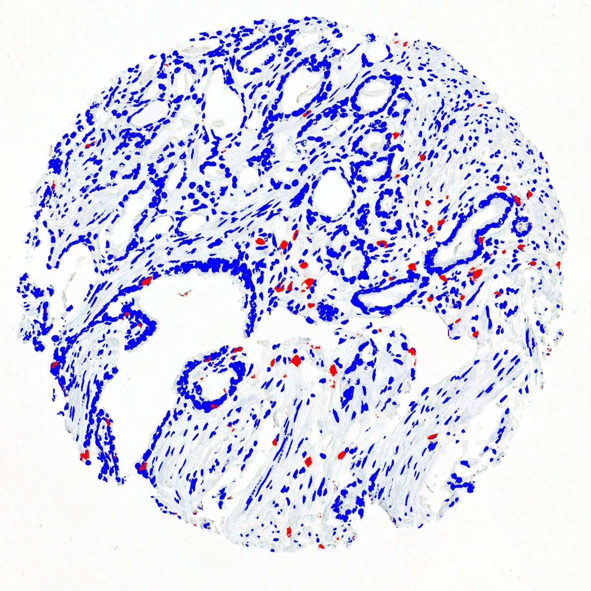
Automated detection of immunohistochemistry labels on a tissue microarray
Having over 30x the storage capacity of a typical high-end imaging workstation, with expandability for more, the optional BioPipeline Server with direct dedicated 10GbE network connection allows for more uninterrupted imaging and offline processing and analysis.
BioPipeline’s data transfer rates from the imaging workstation to the dedicated server exceed most traditional networks by 10-100x by using dedicated 10 GbE connections.