Nikon Instruments Inc. | Americas
- en Change Region
- Global Site
High throughput imaging is a term used to describe automated light microscopy and image analysis of large numbers of samples. It is a useful technique for phenotypic screening assays, evaluating the response of cells, cell-based models, or organisms to various types of perturbagens. When selecting a microscope-based system for high throughput imaging, it is important to not only find a system with the necessary features/capabilities, but also to critically evaluate its reliability and capacity – two factors that have a real-world impact on throughput and efficiency.
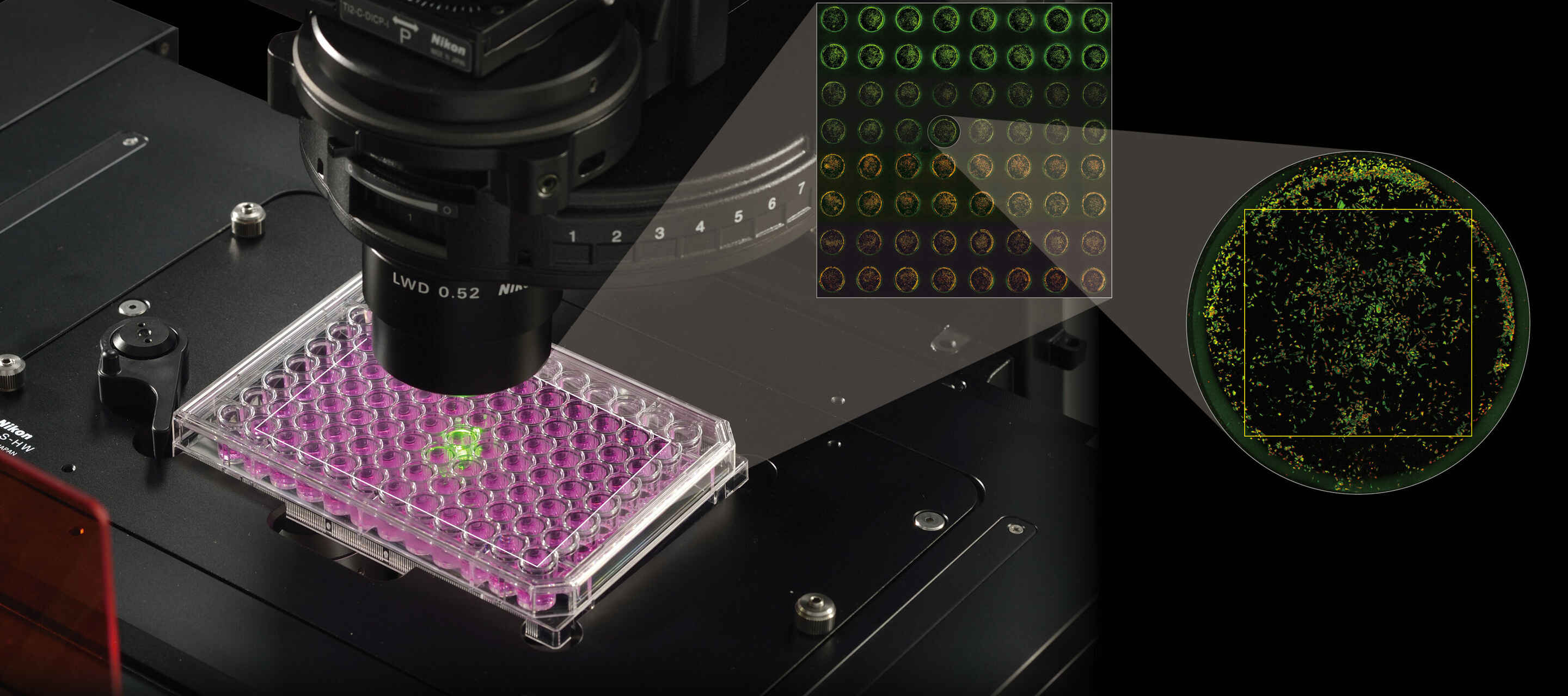
Nikon offers the BioPipeline series of instruments for both high throughput and high content imaging applications. The BioPipeline LIVE high content imaging system is Nikon’s solution for high throughput and high content imaging of live cells kept in vitro. Based on our flagship ECLIPSE Ti2-E motorized inverted microscope, this system features an integrated incubation system and sample exchange robotics, allowing for automatic exchange of up to 44 multi-well plates while maintaining complete environmental coverage.
The BioPipeline PLATE high content imaging system is similar to the BioPipeline LIVE, also based on the Ti2-E microscope but without incubation. Like all BioPipeline systems, the BioPipeline PLATE is completely integrated using the NIS-Elements software, which includes the NIS-Elements Scheduler tool for partitioning time for different automated acquisitions among multiple users and/or samples.
The Nikon BioPipeline SLIDE high content imaging system is instead configured on an ECLIPSE Ni-E upright motorized microscope and intended for applications such as slide scanning/whole slide imaging of fixed samples prepared on standard microscope slides. This system features a Marzhauser Slide Express 2 robotic slide loader, which is able to hold and exchange up to 120 slides. All BioPipeline systems are available with a dedicated server that, with a 10 GbE connection and 200 TB storage space, will help keep your imaging and analysis pipeline from becoming clogged.
Both the BioPipeline LIVE and BioPipeline SLIDE can be outfitted with a confocal system, with our AX / AX R confocal microscopes being applicable towards high throughput imaging, featuring an unmatched 25 mm field of view for a point-scanning system. The AX R additionally features Nikon’s high-speed resonant scanning system, which further accelerates data acquisition.
●: included, ⚬: option
| BioPipeline LIVE High Content Imaging System |
BioPipeline PLATE High Content Imaging System |
BioPipeline SLIDE High Content Imaging System |
|
|---|---|---|---|
| Maximum Sample Capacity | 44 | 44 | 120 |
| Automated Sample Exchange | yes | yes | yes |
| Intended Vessel/Slide Type(s) | 96-well plates 384-well plates |
96-well plates 384-well plates |
Standard glass microscope slides |
| Z Focus Correction | Perfect Focus System 4 (PFS4) Auto-Focus |
Perfect Focus System 4 (PFS4) Auto-Focus |
Auto-Focus |
| Contrast Techniques | BioPipeline LIVE | BioPipeline PLATE | BioPipeline SLIDE |
| Brightfield | yes | yes | yes |
| Phase Contrast* | yes | yes | yes |
| Differential Interference Contrast (DIC) | no | no | yes |
| Darkfield | no | no | yes |
| Nikon Advanced Modulation Contrast (NAMC)* | yes | yes | no |
| Widefield Fluorescence | yes | yes | yes |
| Confocal | yes | yes | no |
*These contrasting techniques may not be well-suited for all applications and magnification ranges due to the meniscus effect within individual wells.
Software dialog during a multi-dimensional acquisition in a well plate.
It is not uncommon for the terms “high throughput” and “high content” to be used interchangeably. The key difference is the dimensionality, or information content, of the acquired image data. Higher information content can be helpful for better characterizing complex phenotypes. “High content” often implies the use of multiple fluorescence channels for characterizing multiple biomarkers. In contrast, high throughput refers to how many samples can be processed rather than the information content of the collected data. It is also possible to perform high content imaging in a high throughput manner (lots of samples, high data dimensionality), an approach sometimes referred to instead as “high content screening.”
Fortunately, Nikon’s BioPipeline systems are suited to both high throughput and high content approaches. Being based on our core microscopes these systems are generally more modular and upgradeable than comparable “box systems” such as plate readers. Cameras, objective lenses, confocal scanners, and more can be swapped in/out on existing systems.
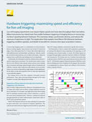
Multicolor widefield fluorescence image of a field of cells
The same image as above following use of the Clarify.ai software module for blur removal.
Many believe that the greatest promise of artificial intelligence in microscopy is the unlocking of new types of image analyses. It is certainly true that new types of analyses are important and useful, a prime example being the prediction of staining patterns in unstained samples (e.g. prediction of nuclear DAPI staining from phase contrast images). However, arguably the greater benefit of AI is the ability to perform common analyses significantly faster than traditional approaches, changing the rules of experiment design.
Nikon is committed to the design of reliable deep learning-based image analysis tools under the umbrella of NIS.ai – a growing series of software modules available within our NIS-Elements software. NIS.ai modules that can impact high throughput imaging include:
There are a variety of confocal microscopy systems on the market, with the two primary design types being the point-scanner and spinning disk. Confocal scanners are desirable when it is important to observe crisp 2D planes within thick specimens. Both the BioPipeline LIVE and BioPipeline PLATE can be configured with these types of confocal systems.
Spinning disk confocal systems are valued for their ease-of-use and imaging speed, but are generally limited in imaging depth to less than 50 μm. Nikon is proud to make available spinning disk systems with a large field of view (FOV) to support high throughput confocal imaging, including the Yokogawa CSU-W1 (~23 mm FOV) and the Crest Optics X-Light V3 (25 mm FOV).
Point-scanning confocal systems can image to hundreds of μm deep in the sample. The Nikon AX / AX R confocal systems provide this capability along with a large 25 mm field of view (the largest commercially available) to support high throughput imaging. Additionally, the AX R uses our next-generation low-noise resonant scanning system to reduce imaging time while maintaining quality.
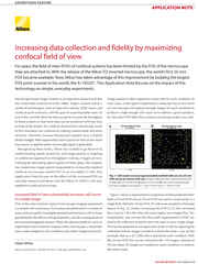
With a conventional 18 mm field of view (FOV), it would take 48 different tiled images to make this large image
With the large 25 mm FOV of the BioPipeline LIVE, BioPipeline PLATE, and AX / AX R confocal systems, only 24 frames would be required at the same magnification.