Nikon Instruments Inc. | Americas
- en Change Region
- Global Site
Electrophysiology is the study of the electrical properties of biological specimens. While this term may refer to classic tissue/organ-scale electrical recording methods used in clinical settings (e.g. electrocardiography), as well as the cellular/sub-cellular level recording methods typically performed in conjunction with an optical microscope in a research lab and of interest here. Patch-clamp recording continues to serve as the standard for recording electrical activity in single neural or muscle cells (or single ion channels). Recording can be performed within a variety of model systems, including in vitro cell cultures, excised tissue, and in vivo.
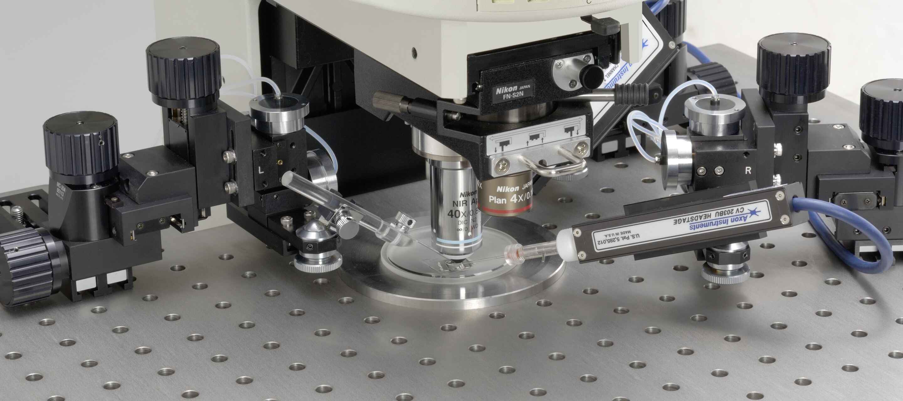
Patch clamp recording methods require suction of a “patch” of the cell membrane using a micrometers-thick glass pipette (positioned using a micromanipulation system). A thin silver wire within the pipette allows for electrical recording. Whole-cell patch and perforated patch methods additionally allow for fluorescent stains (e.g. indicators for voltage, calcium) to be added for microscopic observation, in addition to other experimental agents.
The ECLIPSE FN1 upright microscope is Nikon’s core microscope solution for electrophysiology applications. This microscope features a slim I-shaped frame that prioritizes access to the sample, providing room for positioning of micromanipulators and other equipment. The 2-position front/back-sliding nosepiece allows for switching of objectives, the objective is retracted by 15 mm when switching between positions in order to avoid disturbing the sample and micromanipulation equipment.
The FN1 is compatible with a range of confocal and multiphoton solutions for imaging deeper. The CSU Series spinning disk confocal microscopes from Yokogawa allow fast optical sectioning in live cells and tissue slices. The Nikon AX / AX R confocal microscopes are point-scanning instruments, which allow for deeper imaging than with a spinning disk. The AX R is the resonant-scanning model, and capable of imaging at a rate of up to 720 frames per second (FPS; 2048 x 16 band scan mode), allowing for imaging calcium dynamics and other similarly fast dynamics.
●: included, ⚬: option
| ECLIPSE FN1 Upright Microscope (widefield only) |
Yokogawa CSU-X1 Spinning Disk Confocal* |
Yokogawa CSU-W1 Spinning Disk Confocal* | AX / AX R Confocal System* |
A1 MP+ / A1R HD MP+ Multiphoton System* |
|
|---|---|---|---|---|---|
| Field of View | 25 mm diagonal (widefield; circular) |
10 x 7 mm (confocal; rectangular) |
17 x 16 mm (confocal; rectangular) |
25 mm diagonal (confocal; square) |
18 mm diagonal (multiphoton; square) |
| Zoom Magnification | 0.35X, 2.0X, 4X (widefield; with FN-DP Double Port & Magnification Changer) 1.0X, 1.25X, 1.5X, 2.0X (widefield; with FN-MT Magnification Changer Turret) |
1 – 1000X (confocal; continuously variable scan zoom) | 1 – 1000X (multiphoton; continuously variable scan zoom) | ||
| Relative Imaging Depth Limit | ~ 5 μm ~ 15 – 25 μm (with deconvolution) |
~ 50 μm | ~ 50 – 100 μm | ~ 100 – 500 μm | ~ 1.4 mm (with 1300 nm excitation) |
| Compatible Contrasting Techniques | ECLIPSE FN1 | CSU-X1* | CSU-W1* | AX / AX R* | A1 MP+ / A1R HD MP+ |
| Brightfield | yes | no | no | no | no |
| Confocal - Point-Scanning | no | no | no | yes | yes |
| Confocal - Spinning Disk | no | yes | yes | no | no |
| Differential Interference Contrast (DIC) | yes | no | no | no | no |
| Infrared DIC (IR-DIC) | yes | no | no | no | no |
| Multiphoton - Point Scanning | no | no | no | no | yes |
| Oblique Illumination | yes | no | no | no | no |
| Simple Polarized Light | yes | no | no | no | no |
| Widefield Fluorescence | yes | no | no | no | no |
*Compatibility of a confocal and multiphoton system in this table with additional contrasting techniques is dependent on the microscope stand to which the system is attached. For example, configuring an AX / AX R confocal system on an ECLIPSE FN1 microscope would make it possible to use contrasting techniques that are compatible with the FN1.
IR-DIC image of patch clamped neuron in explanted brain tissue acquired using an FN1 upright microscope.
While fluorescence labeling of samples used in electrophysiology experiments is fairly common, it is not required for patch-clamp recording. Thus, a label-free imaging technique is usually employed for visualizing the sample. This can be challenging as samples are often quite thick (e.g., brain slices that are hundreds of μm thick), scattering light and limiting the depth with which sufficiently detailed observation can be performed.
While label-free techniques such as brightfield and phase contrast microscopy may be useful for thinner samples, such as adherent cell cultures, they are not suited to imaging in thick tissue sections or in vivo. Differential interference contrast (DIC) is an option due to its optical sectioning capability, allowing for deeper imaging while maintaining high resolution (DIC utilizes the full numerical aperture of the objective lens).
Unfortunately, DIC alone is often insufficient. For this reason, Nikon offers near-infrared DIC (IR-DIC), which uses near-infrared (NIR) illumination and compatible optical components. NIR light penetrates further into scattering samples thanks to the longer wavelengths of NIR light compared to visible light, making it possible to identify individual neurons at depths where visible light DIC fails.
Microscopes are usually configured with water dipping objective lenses for electrophysiology experiments – where the tip of the lens is directly immersed in the aqueous sample medium (without cover glass). Water dipping objectives for electrophysiology often have tips made of a chemically inert and electrically insulating material, such as ceramic. The form factor of the objective lens is also important, the tip of the objective should have a steep approach angle towards the tip in order to provide maximum access to the sample for micromanipulators.
The Nikon CFI60 Water Dipping Series objective lenses feature high degrees of aberration correction and transmittance into the NIR, making them suitable for electrophysiology research, especially in combination with IR-DIC or multiphoton imaging. All CFI60 Water Dipping objective lenses are compatible with IR-DIC, with the NIR 40X W and NIR 60X W lenses additionally benefitting from their enhanced NIR transmission. The CFI Plan 100XC W features a correction collar for spherical aberration correction.
The Nikon CFI75 Water Dipping Series objective lenses includes 16X and 25X water dipping lenses, and are physically larger than Nikon’s CFI60 lenses, featuring a thread size of M32 and parfocal distance of 75 mm in order to accommodate the large optics required to collect light at a wide enough angle to achieve a high numerical aperture (NA) together with a longer working distance. The CFI75 LWD 16X W is a popular choice as a “single objective solution,” with magnifications of 5.6X, 32X, and 64X accessible using the FN-DP Double Port & Magnification Changer. The CFI75 Apochromat 25XC W 1300 is one of the brightest objectives Nikon manufactures, and features high transmittance and aberration correction out to 1300 nm. All CFI75 Water Dipping objective lenses are also compatible with IR-DIC.
Optogenetic stimulation of cells expressing optogenetic actuators for various functions, such as the light-gated ion channel channelrhodopsin-2 (ChR2), is a popular method for a variety of experimental applications. Optogenetic electrical stimulation of neural and muscle cells is complemented by patch clamp recording as a readout. This method allows for the local and long-range functional connections of different neurons to be evaluated directly. Optogenetic stimulation is generally accomplished using patterned illumination from a dedicated photostimulation device.
The ECLIPSE FN1 microscope can be configured with a manually-adjustable single-point photostimulation device or digital micromirror device (DMD). DMDs allow for application of arbitrary illumination patterns with near-diffraction limited structure. This allows for precise cellular-to-sub-cellular targeting of stimulation with user-programmable patterns and switching rates of up to 4000 Hz.