Nikon Instruments Inc. | Americas
- en Change Region
- Global Site
Glial cell surrounded by axons in a rat neuronal culture labeled for microtubules and actin
Dr. Christophe Leterrier, NeuroCyto, INP, Marseille, France
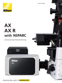
Confocal microscopes have been commercially available now for over 25 years. How can newer iterations of a fundamentally simple instrument continue to innovate? What changes can redefine how a confocal is used, and what data can be collected? Introducing the Nikon AX/AX R Confocal Microscope System, our 10th generation point scanning confocal, giving you more of everything: Leveraging Artificial Intelligence (AI), expanding the number of colors, improving pixel density, sensitivity and speed.
These are significant additions in terms of expanding the range of experiments possible with a point scanning confocal, while increasing the usability and functionality of the instrument, all in a modular and upgradable platform.
Nikon AX is the new standard in confocal imaging.
Whole mouse bladder optically cleared with iDISCO and acquired at 8192 x 8192 pixels using a 2x Plan Apo objective, effective pixel size 0.6 μm (over 5x the spatial resolution of a typical monochrome CMOS camera).
Courtesy of Dr. Gerry Apodaca, Integrative Systems Biology, Department of Medicine, University of Pittsburgh in collaboration with Dr. Alan Watson at the Center for Biological Imaging, University of Pittsburgh.
With the largest field-of-view on both inverted and upright microscope stands available (25mm diagonal), more specimens fit in one FOV than ever before.
Coupled with scanning sizes up to 8k x 8k, sampling beyond the optical diffraction limit is possible even at low magnifications with the AX/AX R.
Using lower magnifications with longer working distances and high numerical apertures enables more flexible specimen preparations to be used, while the large FOV allows simultaneous high resolution in one image. Drive research and drug discovery with more data in every image, collected at higher speeds.

The AX/AX R has a 25mm diagonal FOV, much larger than other confocal instruments.
Danio sp. 2d+ embryo 4X*
Cleared adult mouse brain acquired with 1x objective in one acquisition of one FOV*
Drosophila sp. Embryo development easily fits within the FOV using a high NA 25x SIL 1.05 NA objective*
* It would not be possible to capture this sample in one FOV or at this resolution with other commercial confocal systems
Laser scanning confocal imaging is principally challenging on specimen viability, as it applies focused laser illumination point by point on a sample.
The AX R’s high speed resonant scanning, which decreases the illumination time by more than 20x typical confocal scanning times, greatly reduces biases caused by merely acquiring images. Reducing the acquisition time also allows for extremely high-speed imaging (up to 720 fps @ 2048 x 16).
The result: longer time-lapse imaging at better frame rates with living samples, enabling the detection of dynamic events. This is useful for imaging living samples, and dramatically increases the efficiency of drug discovery research and multipoint assays.
Time-lapse Z series maximum intensity projection images of a developing Drosophila embryo expressing PLC-PH::GFP (PIP2) acquired every 10 minutes for 12 hours at 2K x 1K pixels using a 25x silicone immersion objective.
Courtesy of Yang Hong Laboratory, Department of Cell Biology, University of Pittsburgh in collaboration with the Center for Biological Imaging.
Confocal imaging, notoriously slow because of its point-scanning requirement for high quality 3-dimensional imaging at high resolution, is greatly changed by fast imaging with the AX R’s resonant scanning capabilities.
Utilizing 2048 x2048 pixel resonant scanning and a 25mm FOV on a large intestinal sample montage, acquiring 25 high-resolution images and merging them in under 2 minutes.
With a full 25mm FOV, up to 8192 x 8192 pixels, incredibly low noise and the capability for supravideo frame rates, the AX/AX R allows for spectacular imaging with high resolution at any magnification.
This capability benefits the entire range of imaging applications from capturing the details of target selection in drug discovery to whole organisms and system biology
Mouse muscle acquired with a 25x SIL immersion objective using 2048 x 2048 pixel resonant scanning
Maximum intensity projection of Z stack images of marmoset brain acquired with a 60x 1.27 NA water immersion objective using 2048 x 2048 pixel resonant scanning and a DUX-VB detector with user-defined emission bands.
The AX/AX R’s all new DUX-VB detector custom-tunes emission bandwidths to a library of labels and probes, and provides the freedom to fine-tune emission bands to minimize unwanted fluorescence.
Simply select the number of labels in your specimen and their catalog names. Alternatively, you can define the desired emission ranges, or even simply the emission color: the AX/AX R and NIS-Elements software does the rest, including optimizing the dichroic mirror and laser excitation choices best suited for imaging. Or, acquire hyperspectral images in up to 66 emission channels for unmixing.
Optionally, the AX/AX R’s base DUX-ST detector allows up to 12 discreet bandpasses of emission, upgradable to 18.
And all detector systems can be customized with high sensitivity and low noise GaAsP or Multi-alkali PMT detectors to provide the best detector for sensitivity and wavelength response requirements as well as budgets.
Controlling the manufacturing and implementation of optics from raw materials all the way to complete microscope systems brings unparalleled optical quality and performance.
Complementary optical design means the confocal system, microscope, and objectives all are optimized and matched for superior quality and resolution.
Nikon’s CFI60 and 75 infinity corrected optical system has numerous options for magnifications, working distances, and immersion mediums, paired for use with an extremely wide variety of samples and specimen preparations.
A software-controlled, automatic water dispenser enables long-term time-lapse imaging using refractive-index matching water immersion objectives in any environment, including incubation.
Moves the objective correction collar to the optimum position for best resolution both remotely and by software control. Motorized collars allow users to adjust the correction collar without disturbing the specimen position, even in incubated enclosures or environmental chambers.
The Ti2-E microscope supports up to 5 episcopic illumination sources, which can be used in tandem with AX/AX R confocal imaging: total internal reflection fluorescence (TIRF), point, raster or field stimulation devices, and fluorescence light sources can all be integrated onto the same microscope stand, and used in the same experiments.
The incident angle of a laser and corresponding penetration depth of the evanescent field can be controlled via NIS-Elements software. When multiple TIRF modules are mounted, the penetration depth can be independently set for each wavelength.
The XY galvano scanning unit can stimulate the desired area of a sample using laser point scanning. It allows simultaneous photostimulation and confocal imaging.
The DMD module enables photoactivation of user-specified patterns rather than photoactivation of a single spot. This allows stimulation of multiple points and tracking of their behavior. The DMD module can be used with either laser illumination or less phototoxic LED illumination.
This device enables photostimulation over a 400 to 700 nm wavelength range*, allowing photostimulation and imaging with visible light. Simultaneous stimulation, sequential stimulation and manual stimulation are available.
*Depends on filter cube type.