Products and Promotions may differ based on your selected Region.
Return to the top of the page
Eingestellte Produkte
Nikon BioImaging Labs provide contract research services for microscope-based imaging and analysis to the biotech, pharma, and larger research communities. Each lab's full-service capabilities include access to cutting-edge microscopy instrumentation and software, but also the services of expert biologists and microscopists, who are available to provide quality cell culture, sample preparation, data acquisition, and data analysis services.
Tragen Sie sich hier in unsere Mailingliste ein, um als Erster über unsere neuen Produkte und exklusiven Angebote informiert zu werden!
Im Nikon Instruments Learning Center finden Sie eine Vielzahl an interaktiven Tutorials, in denen Inhalte zu Grundlagen- bis zu fortgeschrittenen Themen behandelt und erklärt werden.
Schritt-für-Schritt-Anleitungen zum Justieren und Bedienen ausgewählter Nikon-Mikroskope.
Search and filter over 125,000 Open Access Articles that utilize Nikon products and supported third party systems.
Finden Sie das richtige Nikon-Objektiv für Ihre Fragestellungen.
Laden Sie Software- und Firmware-Programme für Nikon-Mikroskopprodukte herunter.
Nikons MicroscopyU ist eine Top-Adresse für Ausbildungs- und Informationsmaterial über die optische Mikroskopie.
Die Nikon Small World-Website – alles über Fotomikrografie-/Videowettbewerbe.
November 2025
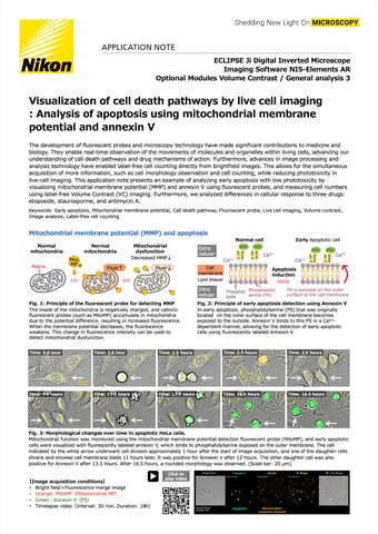
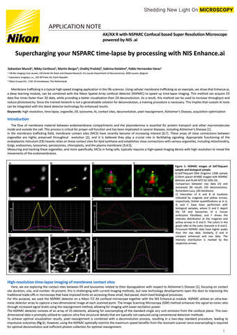
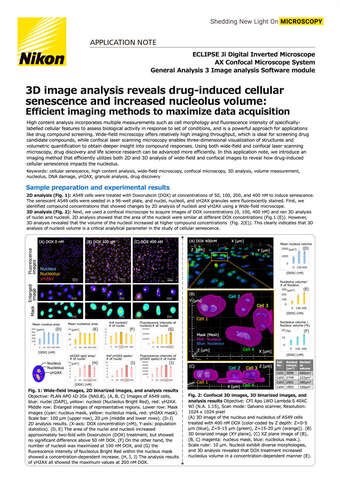
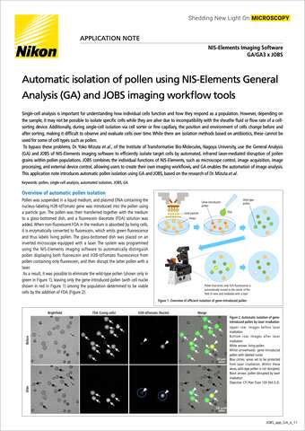
Weiterlesen und Download
Juli 2025
November 2024
November 2023
März 2021
Januar 2021
September 2020
Januar 2020
Dezember 2017
Download 4.43MB