Nikon Instruments Inc. | Americas
- en Change Region
- Global Site
April 2023
In vitro human Blood-Brain Barrier (BBB) models are essential to assess the neurotherapeutic transport efficacy in pre-clinical studies and to understand the pathological neurovascular functions in various diseases. This application note introduces an example of building a robust and cost-effective in vitro BBB model using AIM Biotech’s 3D cell culture chips. Through the co-culture of human brain microvascular endothelial cells (EC), human brain pericytes (PC) and human astrocytes (AC), a microvascular network within a fibrin gel was self-organized. A Nikon confocal microscope contributed to monitor physiologically relevant structures in the 3D BBB model stained by cell-specific and BBB-specific markers. Also, quantitative image analysis was used to compare its permeability to in vivo values measured in rat brains.
Fig. 1: Top view and cross section diagrams of a site in the idenTx 40 system
By utilizing AIM Biotech’s idenTx 40 (Fig. 1), 3 types of cells were co-cultured for 7 days under the condition described in Table 1. The tri-culture yields a
perfusable 3D microvascular network within the chip through the vasculogenesis process (Fig. 2). Widefield microscopy was used to measure its permeability
and confocal microscopy was used to verify the marker signals and 3D structures of each cell.
Fig. 2: A schematic diagram of a self-organized 3D human BBB in an idenTx 40
| Cell | Seeding concentration |
|---|---|
| Human Brain Microvascular Endothelial Cells (cAP-0002): Angio-Proteomie | 6 M/ml |
| Primary Human Brain Pericytes (ACBRI 498): Cell Systems | 0.5 M/ml |
| Human Astrocytes (Catalog #1800): ScienCell | 1 M/ml |
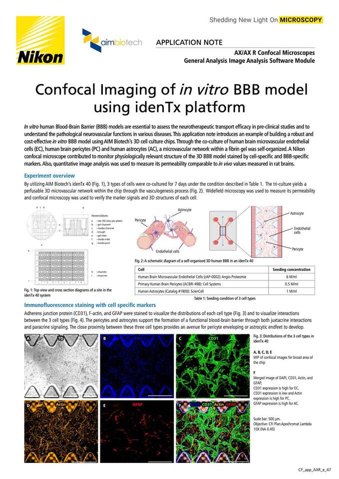
Adherens junction protein (CD31), F-actin, and GFAP were stained to visualize the distributions of each cell type (Fig. 3) and to visualize interactions
between the 3 cell types (Fig. 4). The pericytes and astrocytes support the formation of a functional blood-brain barrier through both juxtacrine interactions
and paracrine signaling. The close proximity between these three cell types provides an avenue for pericyte enveloping or astrocytic endfeet to develop.
Fig. 3: Distributions of the 3 cell types in
idenTx 40
A,B,C,D,E
MIP of confocal images for broad area of
the chip
F
Merged image of DAPI, CD31, Actin, and
GFAP, CD31 expression is high for EC.
CD31 expression is low and Actin
expression is high for PC.
GFAP expression is high for AC.
Scale bar: 500 μm.
Objective: CFI Plan Apochromat Lambda
10X (NA 0.45)
Fig. 4: Interactions between the 3 cell types in idenTx 40
A,B,C
A is a 3D rendered image of a microvascular network that shows complex interactions between the vasculature, pericytes and astrocytes.
The area where a pericyte cell envelopes the microvasculature is magnified in B. The area where the astrocytes interact with the microvasculature is magnified in C.
D
A section image of the microvascular network
An EC tubule structure is enveloped by PC (white arrow). Scale bar: 100 μm.
Objective: CFI Apochromat LWD Lambda S 20XC WI (NA 0.95)
The 3D BBB model formed in the idenTx platform also exhibits phenotypes of a healthy BBB through the expression of BBB-specific tight junction proteins
such as CLAUDIN-5 and ZO-1. A basal membrane protein such as laminin is expressed throughout the BBB vascular network, which is another indication of
a functional in vitro BBB.
Fig. 5: Expression of BBB specific markers
A, B, C, D: MIP of confocal images of a broad area of the chip. Scale bar: 500 μm.
a, b, c, d: Magnified images of A, B, C and D. Scale bar: 100 μm.
Objective: CFI Plan Apochromat Lambda 10X (NA 0.45)
In order to quantify microvascular permeability, 70 kDa FITC-dextran was used to perfuse the microvasculature in the idenTx platform. Then time- point imaging was done to assess the fluorescent intensity in the surrounding matrix, which changes over time, to calculate the permeability of 70 kDa dextran across the BBB. (Please refer to the BBB protocol on the AIM Biotech website, where the steps of quantification are shown in detail.)
Fig. 6: BBB Permeability assessment using fluorescent-based imaging
A: Time-point imaging of a BBB microvasculature perfused with 70 kDa FITC-dextran
B: The projected images (top) of the selected areas of the perfused vasculature were binarized (bottom) to distinguish the vasculature from the surrounding matrix.
C: Typical permeability of a 70 kDa fluorescent particle is in the range of 10-7 (cm/s) which is similar to the value obtained from in vivo rat brains.
Quantification of the microvasculature structure is significant for accurate measurement of permeability experiments and consideration of appropriate culture
conditions. General Analysis (GA) in NIS-Elements software enables measurements such as diameter, circularity, and branching point for projected 2D images
to be performed. In addition, GA enables 3D binary masks of vessel structures and medium channel regions to be created from confocal Z stack images (Fig.7)
and accurate 3D measurements such as volume and surface area to be performed.
Fig. 7: Detection of 3D structure from Z confocal stack images
A: 3D image of cell specific markers, B: 3D image of CD31, C: 3D binary mask of vessel (yellow) and medium channel region (blue) Objective: CFI Plan Apochromat Lambda 10X (NA 0.45)
idenTx 40 Plate (AIM Biotech)
Harness AIM’s sophisticated organ on a chip technology and unbeatable ease-of-use, now in a standard SBS plate format that enables 40 simultaneous experiments on a single plate.
The idenTx 40 Plate is all about increasing throughput for critical drug discovery assays that require accurate recreation of human microphysiology, all while working seamlessly with the downstream analytical instruments you already have.
These microscopes achieve high resolution images of 8K x
8K pixels, which is four times that of conventional models.
A large FOV with a diagonal of 25 mm allows acquisition
of a large area of samples in a single scan, reducing
phototoxicity. The AX R’s resonant scanner achieves a high
resolution of 2K x 2K pixels, allowing acquisition of live sample dynamics with
high-speed imaging of up to 720 fps
(2048 x 16 pixels).


