Melike Lakadamyali, Ph.D.
The advanced Fluorescence Imaging and Biophysics Group, ICFO-Institute of Photonic Sciences
関連製品

Please tell us about your research.
My research is focused on studying a central question in cell biology: how does the organization of proteins in space and in time impact cellular function. I study this central question in the context of a number of different biological problems ranging from intracellular transport, chromatin organization to synaptic organization. In all of these cases, I use super-resolution fluorescence microscopy and other single molecule based methods such as single particle tracking, coupled with quantitative analysis, to extract information on the dynamics, stoichiometry and organization of multi-protein complexes and link this information to their function in the cell. Overall, I strive to address fundamental questions in biology, which also have implications for health and disease.
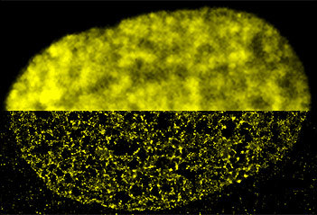
What applications do you use the Nikon N-STORM systems for?
I am using the Nikon N-STORM system mainly for super-resolution imaging. In particular, in the past several years, in collaboration with Maria Pia Cosma at the Center for Genomic Regulation (CRG), Barcelona, we have been imaging the organization of nucleosomes inside the nucleus of a number of different cell types using the N-STORM system. These studies allowed us to gain new insights into how chromatin is organized at the nanometer length scales, and find and important link between this organization and cell state.
We found that nucleosomes are organized in groups, which we named nucleosome clutches cannot be visualized with the conventional microscope. The size of the nucleosome clutches is cell specific and correlates with pluripotency. We published all this in the Cell paper.
 Ricci, M.A., Manzo, C. Garcia-Parajo, M.F.Lakadamyali, M., Cosma, M.P., Cell, 2015
What do you value about this Nikon system?
The best part of the Nikon system is the fact that it is user friendly. I am a physicist but my work is focused on biophysics and I work with biologists on a regular basis. Most biologists are not very comfortable using custom-built microscopes that occupy an entire optical table and have many manual components that must be modified or adjusted during experiments. A physicist with hands-on experience with optical setups can easily use and manipulate these setups but most biologists are looking for easy to use, turn-key systems that are robust and reliable. The Nikon system makes it much easier for me to collaborate with biologists.
What are the future demands for microscopes and imaging?
Light microscopy has undergone a revolution in the past decade. We have seen that it is possible to overcome one of the fundamental limitations in physics, the diffraction limit, by using clever tricks with the fluorescent on and off states of fluorophores. This has really opened new doors into understanding biology at the smallest length scales. I believe the immediate future will see an explosion of new discoveries in biology as these methods will go from a specialist’s tool to everyday use. However, we still need to solve major challenges, mainly the ability to image deep inside thick samples, the ability to image fast dynamic processes in living cells with nanoscale spatial resolution and the ability to extract meaningful quantitative information from the super-resolution images. As we push the biological applications of super-resolution methods, we will need to overcome these challenges and the future demands will mainly be in these topics.