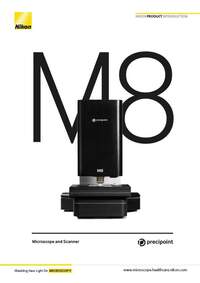
PreciPoint Microscope and Slide Scanners
Products and Promotions may differ based on your selected Region.
Return to the top of the page
Discontinued Products
Nikon BioImaging Labs provide contract research services for microscope-based imaging and analysis to the biotech, pharma, and larger research communities. Each lab's full-service capabilities include access to cutting-edge microscopy instrumentation and software, but also the services of expert biologists and microscopists, who are available to provide quality cell culture, sample preparation, data acquisition, and data analysis services.
Sign up to our mailing list to be first to hear about our new products and exclusive offers!
Free interactive microscopy e-Learning courses, including product-specific courses.
Step-by-step guides for aligning and operating select Nikon microscopes.
Find the right Nikon objective to fit your workflow.
Download software and firmware programs for Nikon microscope products.
Search and filter over 125,000 Open Access Articles that utilize Nikon products and supported third party systems.
Input appropriate parameters to calculate important performance specifications such as resolution, depth of field, sampling rate, confocality, and more.
Nikon's MicroscopyU is a top source for educational information about optical microscopy.
Operating modes tailored to diverse workflows
Live-mode, InstantScan mode, SlideScan-mode
Light
Transmitted Light; LED, brightfield
Barcode scanning
Upon request
Supported objectives
20x to 60x air
20x to 100x air and oil
Dimensions
45 cm x 40 cm x 30 cm; 25 kg
Automated Microscope
X-Y-stage automated, Z-axis automated
Handling
Controlled with computer; Live remote control due to automation
Scanning parameters
Whole Slide Imaging or partial digitization
Scanning algorithms
Different scanning algorithms tailored to different slide qualities
InstantScan-mode
Large field of view within seconds at high resolution
Scanning speed per slide
20x: 2 min
100x: 1 hr20x: 2 min
Scanning resolution per slide
60x: 0.85 NA: 0.16 μm/px40x: 0.75 NA: 0.24 μm/px20x: 0.50 NA: 0.479 μm/px
100x: 1.3 NA: 0.096 μm/px60x: 0.85 NA: 0.160 μm/px40x: 0.75 NA: 0.240 μm/px20x: 0.5 NA: 0.479 μm/px
Slide capacity
25 x 75 mm (2 slides) or 50 x 75 mm (1 slide)
Z-Axis Resolution
25 nm
XY-Axis Resolution
78 nm
Z-Stacking
Yes
Operating software
MicroPoint included
Viewer software
ViewPoint* included (unlimited users)
Storage
PreciCloud slide storage
Image analysis
Several software applications on request, based on customer needs
Computers
Various computers recommended and approved by PreciPoint
Microscope computer connection
USB 3.0
Image output formats
PNG, JPEG, TIFF, BMP, VMIC, XLS, and many more
Download 608KB