Nikon Europe B.V. | Europe & Africa
- en Change Region
- Global Site
Spatial omics represents a transformative evolution in the life sciences – enabling researchers to map RNA expression, protein abundance, and the molecular interactions between them within tissue architecture. By preserving spatial context, these approaches reveal how cellular identity and function are shaped by microenvironmental cues, tissue organization, and disease‑related changes. This transition from bulk or dissociated single‑cell methods to spatially resolved analysis provides unprecedented insight into cellular neighborhoods, lineage trajectories, and pathological microstructures.
A significant branch of spatial omics relies on advanced imaging modalities capable of detecting dozens – or even hundreds – of molecular targets across large tissue regions. Techniques such as multiplexed fluorescence imaging, cyclic immunofluorescence, in situ sequencing by imaging, and spectral unmixing workflows demand stable illumination, high‑efficiency optics, and robust repeatability. These imaging platforms must balance sensitivity with throughput, ensuring that weak biomarker signals remain detectable while accommodating iterative labeling cycles.
The ECLIPSE Ti2 inverted microscope provides a large field of view (FN 25), maximizing the capture area of modern CMOS sensors and substantially improving acquisition throughput. Its optical stability and compatibility with advanced imaging modes make it a strong foundation for high‑content, multi‑round spatial assays.
The ECLIPSE Ni‑E upright microscope is designed for demanding research workflows requiring motorization, automation, and, at the same time, flexibility. It supports a broad range of advanced applications – ideal for complex multi‑modal spatial omics imaging pipelines that integrate high resolution brightfield and fluorescence scanning.
Immune cells and endothelium in mouse lymph node, acquired by Kromnigon AB
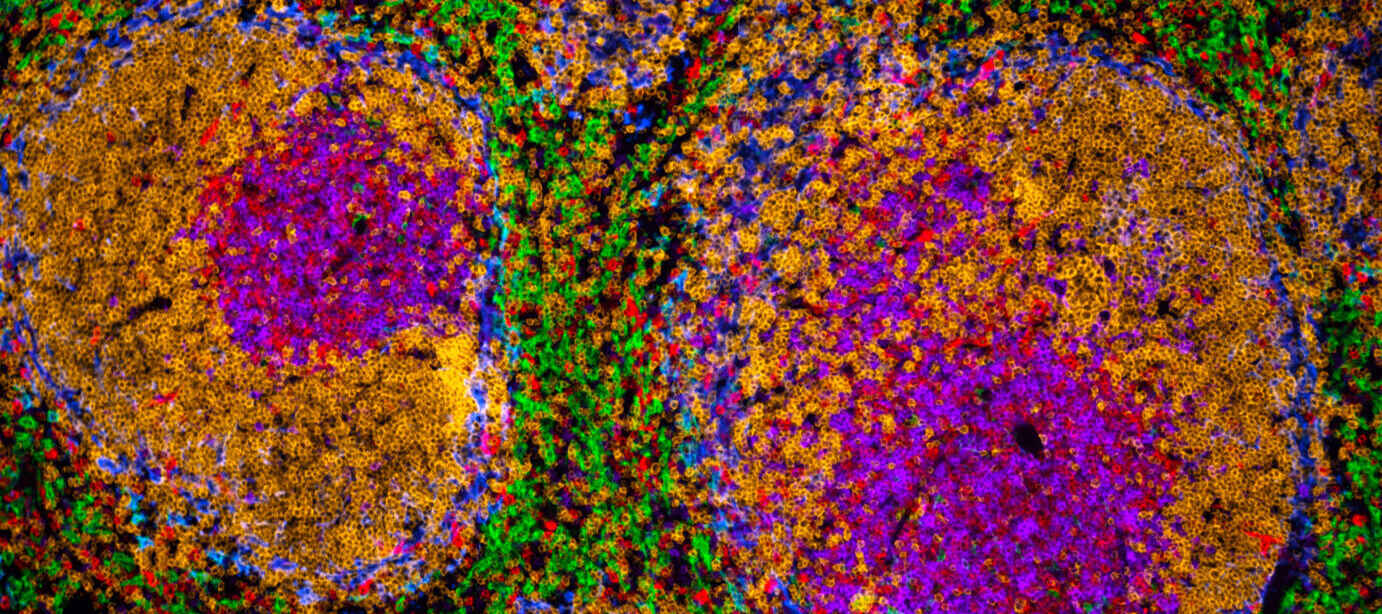
Immune cells in mouse spleen multistained with tyramide signal amplification, acquired by Kromnigon AB
Multi stained mouse intestine with tyramide signal amplification, acquired by Kromnigon AB
Most spatial omics datasets require the imaging of entire tissue sections, organoids, or large biopsy regions. High‑fidelity stitching ensures seamless reconstruction of these expansive fields without loss of registration accuracy, which is essential for downstream quantification of tissue morphology, region‑specific molecular signatures, and cellular localization patterns. High‑throughput acquisition also minimizes photobleaching and sample drift over long acquisition sessions – critical considerations when conducting dozens of imaging cycles.
With its expansive field of view the ECLIPSE Ti2 increases data throughput – reducing the number of required tiles and enabling faster whole‑region imaging for stitched reconstructions. Furthermore, the combination of the ECLIPSE Ti2 hardware with NIS-Elements JOBS allow automated workflows to acquire large amounts of data in an autonomous fashion. It can even integrate General Analysis pipelines for selecting Regions of Interest for further imaging at higher resolution, or other smart microscopy applications.
The Slide Scanner’s motorized control and sophisticated piezo scanning capabilities support precise, repeatable tile scanning and large‑area image stitching across full tissue sections of up to 8 slides.
Please contact your local representative for product availability.
February 2026
Multi stained mouse intestine, acquired by Kromnigon AB
Spatial omics workflows often rely on multiplexed imaging capable of detecting many biomarkers in situ. This requires efficient fluorophore excitation, low background signal, and consistent optical performance across repeated cycles of staining, imaging, and stripping. High multiplexing enhances the ability to classify cell types, identify cellular neighborhoods, quantify disease states, and map interactions across tissue landscapes.
The ECLIPSE Ti2 platform’s optimized optics are compatible with high performance scientific cameras. These enable sensitive detection of low‑intensity fluorophores and high‑resolution capture of densely multiplexed samples. Furthermore, JOBs can manage the integration of fluidic devices. Making iterative fluorescence imaging more efficient, automated and reproducible.
The ECLIPSE Ni‑E’s compatibility with advanced fluorescence gives researchers the ability to configure multi‑plex imaging workflows.