Nikon Instruments Inc. | Americas
- en Change Region
- Global Site

November 2021
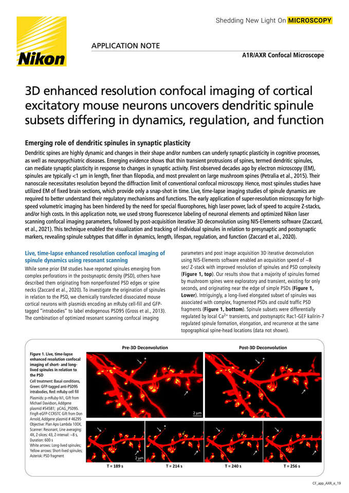
In this application note, we used strong fluorescence labeling of neuronal elements and optimized Nikon laser scanning confocal imaging parameters, followed by post-acquisition iterative 3D deconvolution using NIS-Elements software (Zaccard, et al., 2021). This technique enabled the visualization and tracking of individual spinules in relation to presynaptic and postsynaptic markers, revealing spinule subtypes that differ in dynamics, length, lifespan, regulation, and function (Zaccard et al., 2020).