株式会社ニコンソリューションズ | Japan
Artboard 1
ja Change Region

Global Site

University of Michigan

お問い合わせ

センターオブエクセレンス

University of Michigan

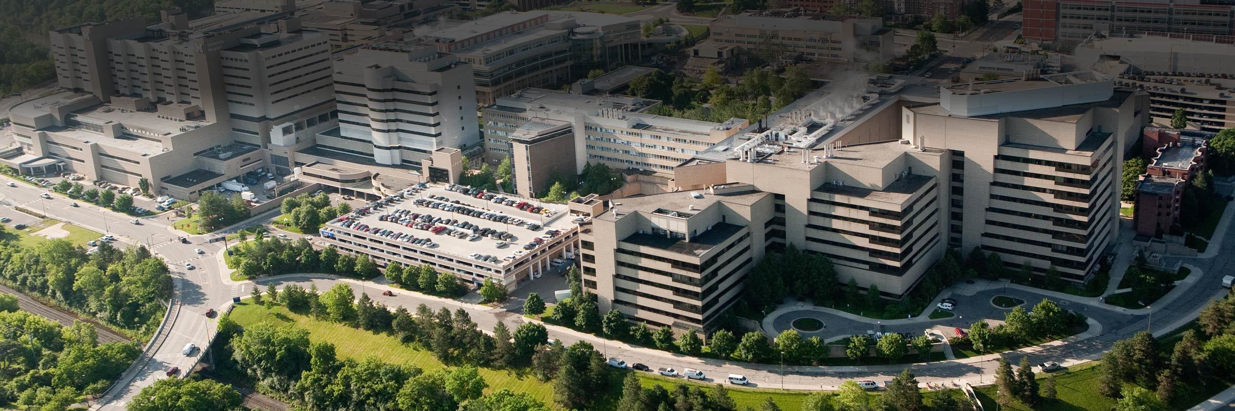
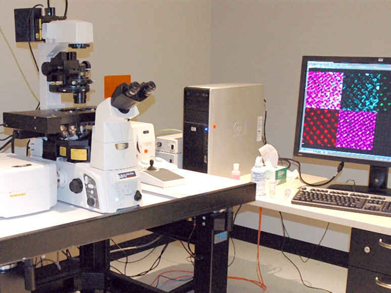
The Microscopy Core provides access to advanced microscopes as well as the expertise of dedicated imaging scientists. We also provide custom image analysis, comprehensive sample preparation for electron microscopy, tissue clearing, and expansion microscopy.

Opening: Tuesday, February 12, 2019; From left to right: Aaron Taylor (Managing Director, Microscopy Core, University of Michigan Medical School), Bob Johnson (Procurement Supervisor, University of Michigan), Andy Davis (General Manager, Sales, Nikon Instruments Inc.), Ben Allen (Microscopy Core Faculty Director, Associate Professor, Cell & Developmental Biology, University of Michigan Medical School), Adiv Johnson (Advanced Biosystems Manager, Nikon Instruments Inc.)

お問い合わせ

CofE Director

email hidden; JavaScript is required

住所

109 Zina Pitcher Drive
Ann Arbor, MI 48105

ウェブサイト

University of Michigan


Systems Available

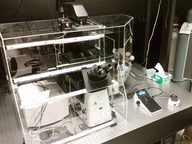
N-SIM/A1R HD Super-Resolution & Confocal System

This system combines super-resolution using the principle of structured illumination and high-speed resonant scanning confocal on a single microscope, making it easy to optimize for resolution or speed with a variety of samples.

Components

  • Ti2-E inverted microscope with Perfect Focus System 4 (PFS4)

  • N-SIM super-resolution structured illumination microscopy system
  • A1R HD high-definition resonant scanning confocal system
  • LUN-V 6-line laser unit (405nm, 445nm, 488nm, 515nm, 561nm, 647nm)
  • DU4G GaAsP 4-channel confocal detector
  • DUV-B 2-channel spectral GaAsP detector
  • Hamamatsu ORCA-Flash4.0 V2+ sCMOS camera
  • Lumencor SPECTRA X light engine
  • Mad City Labs Nano-Z series Z-axis piezo stage
  • Tokai Hit stage-top incubation system
  • NIS-Elements software

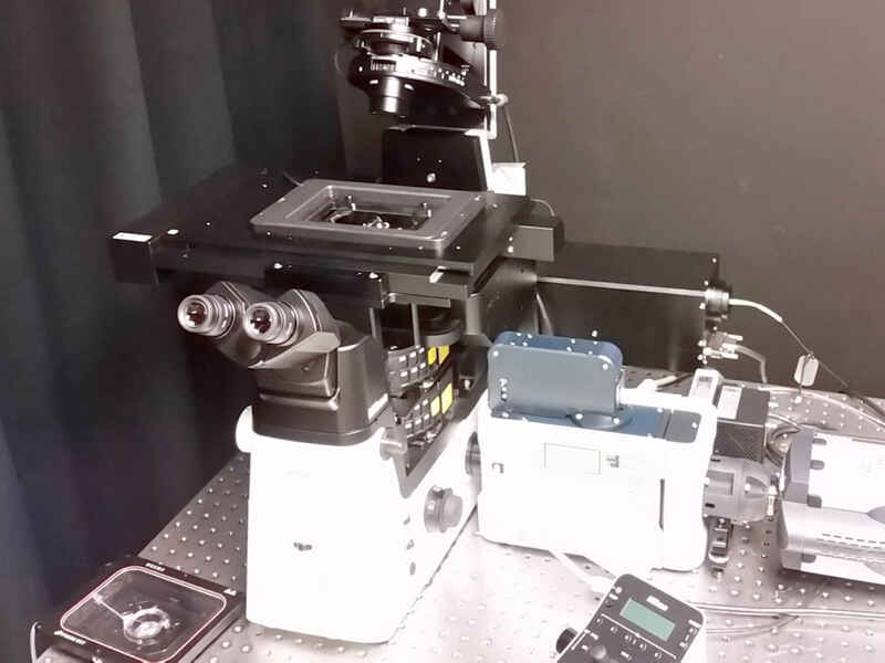
TIRF/FRAP/CSU-X1 Spinning Disk Confocal System

This flexible live cell imaging system is configured on a Ti2-E inverted microscope and features TIRF, spinning disk confocal, and a DMD for patterned photostimulation.

Components

  • Ti2-E inverted microscope with Perfect Focus System 4 (PFS4)

  • Yokogawa CSU-X1 spinning disk confocal
  • H-TIRF motorized illuminator for laser TIRF imaging
  • Mightex Polygon400 DMD for patterned illumination (Lumencor AURA II light engine)
  • LUNF 3-line laser unit (488nm, 561nm, 640nm) for TIRF imaging
  • LUNF 4-line laser unit (405nm, 488nm, 561nm, 640nm) for CSU-X1
  • Hamamatsu W-VIEW GEMINI-2C image splitting optics
  • 2x Andor iXon 888 EMCCD camera
  • Mad City Labs Nano-Z series Z-axis piezo stage
  • Tokai Hit stage-top incubation system
  • NIS-Elements software

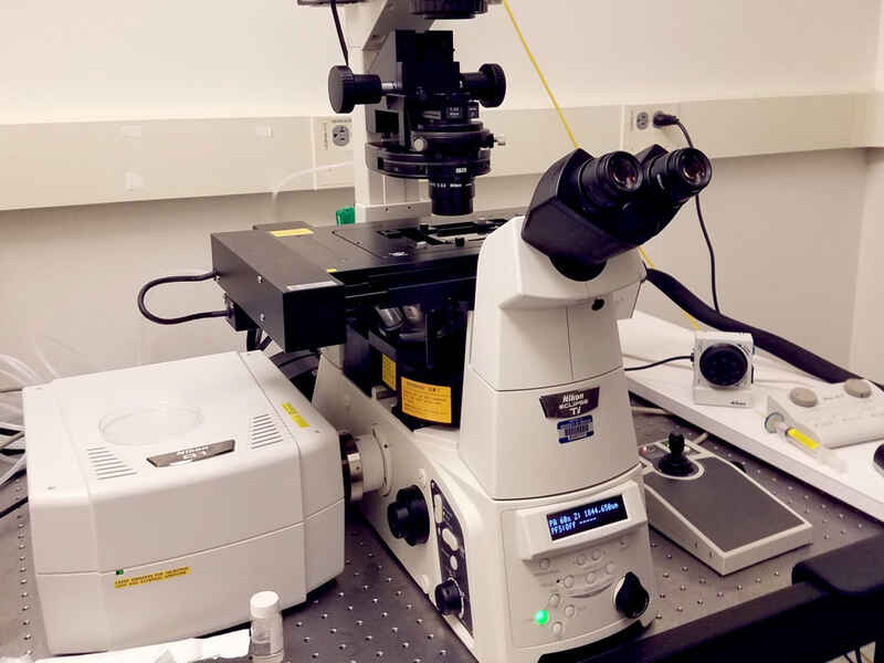
This A1 point scanning confocal system is configured on a Ti-E inverted microscope and well suited to a variety of both live and fixed cell imaging applications.

Components

  • Ti-E inverted microscope with Perfect Focus System (PFS)
  • LU4A 4-line laser unit (405nm, 488nm, 561nm, 647nm)
  • DU4G GaAsP 4-channel confocal detector
  • Tokai Hit stage-top incubation system
  • NIS-Elements software

This A1 point scanning confocal system is configured on a Ti-E inverted microscope and well suited to a variety of both live and fixed cell imaging applications.

Components

  • Ti-E inverted microscope with Perfect Focus System (PFS)
  • LU4A 4-line laser unit (405nm, 488nm, 561nm, 647nm)
  • DU4 4-channel confocal detector
  • NIS-Elements software

A1si Point Scanning Confocal System

This A1 point scanning confocal system is configured on a Ti-E inverted microscope and also features a spectral detector for linear unmixing of signals from multiple emitters.

Components

  • Ti-E inverted microscope with Perfect Focus System (PFS)
  • A1si point scanning confocal system
  • A1-DUS 32-channel spectral detector
  • LU4A 4-line laser unit (405nm, 488nm, 561nm, 647nm)
  • DU4 4-channel confocal detector
  • NIS-Elements software