Nikon Instruments Inc. | Americas
- en Change Region
- Global Site
Center of Excellence
The fundamental goal of the Washington University Center for Cellular Imaging (WUCCI) is to provide access to an integrated infrastructure of light and charged particle based cellular imaging technologies. The Center Director, and his staff provide both professional guidance and work collaboratively with Washington University researchers in assay design, sample preparation and data analysis as well as develop and apply new imaging approaches and informatics methods. The Nikon Center of Excellence within the WUCCI enables investigators to gain unprecedented insights into the dynamic behavior of single molecules and the spatial organization of cells and tissues using state-of-the-art confocal, live-cell and super-resolution microscopies, creating exciting opportunities for discovery in a broad range of basic and translational research aimed at advancing our understanding of human health and disease.
email hidden; JavaScript is required
email hidden; JavaScript is required
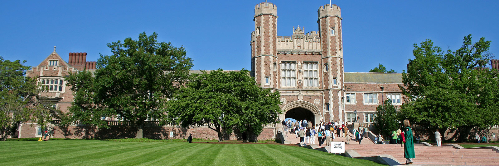
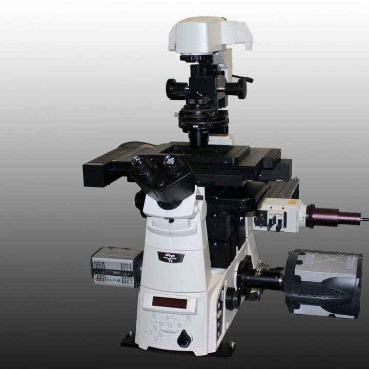
This A1Rsi resonant scanning confocal system is configured on a Ti-E inverted microscope and features a spectral detector for linear unmixing of multiple overlapping signals.
NIS-Elements software
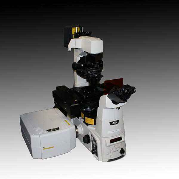
This live cell imaging system features a Yokogawa CSU-X1 spinning disk confocal scanner on Ti-E inverted microscope, as well as emission splitting optics for simultaneous two-color imaging and photostimulation devices for applications such as optogenetics.
Yokogawa CSU-X1 spinning disk confocal
NIS-Elements software
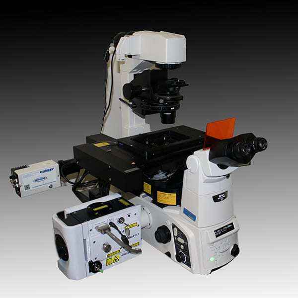
This system features the N-SIM super-resolution structured illumination microscopy system on a Ti-E inverted microscope, suitable for both live and fixed cell imaging.
NIS-Elements software
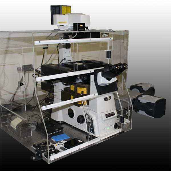
The N-STORM provides super-resolution single molecule localization imaging on the Ti-E inverted microscope, and also good for TIRF imaging.
N-STORM super-resolution single molecule localization microscopy system
NIS-Elements software