Nikon Europe B.V. | Europe & Africa
- en Change Region
- Global Site
March 2023
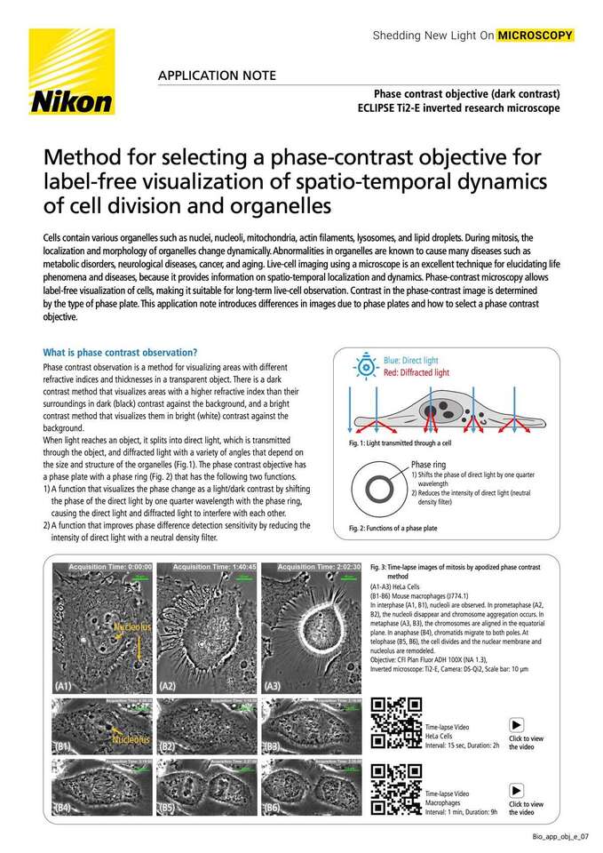
Cells contain various organelles such as nuclei, nucleoli, mitochondria, actin filaments, lysosomes, and lipid droplets. During mitosis, the localization and morphology of organelles change dynamically. Abnormalities in organelles are known to cause many diseases such as metabolic disorders, neurological diseases, cancer, and aging. Live-cell imaging using a microscope is an excellent technique for elucidating life phenomena and diseases, because it provides information on spatio-temporal localization and dynamics. Phase-contrast microscopy allows label-free visualization of cells, making it suitable for long-term live-cell observation. Contrast in the phase-contrast image is determined by the type of phase plate. This application note introduces differences in images due to phase plates and how to select a phase contrast objective.