Nikon Europe B.V. | Europe & Africa
- en Change Region
- Global Site
September 2023
A spindle is a very important structure that is formed to distribute chromosomes to daughter cells during cell division. It is usually located on the inside of the immediate vicinity of the polar body but may also be far from the polar body. Therefore, it is possible that the spindle may be damaged in intracytoplasmic sperm injection (ICSI), which is performed without observation of the spindle, and the fertilized egg may not grow normally. There are also reports of the fertilization rate changing depending on the position of the spindle[1]. Furthermore, it is considered that the presence/absence, position and morphology of the spindle are indicators of oocyte maturation[2]. Therefore, it is very important to visualize the spindle, but it is difficult to observe with a normal microscope.
This application note introduces a spindle observation system that enables spindle identification. By attaching this system to an inverted microscope, ICSI can be performed at the right time while preventing damage to the spindle.
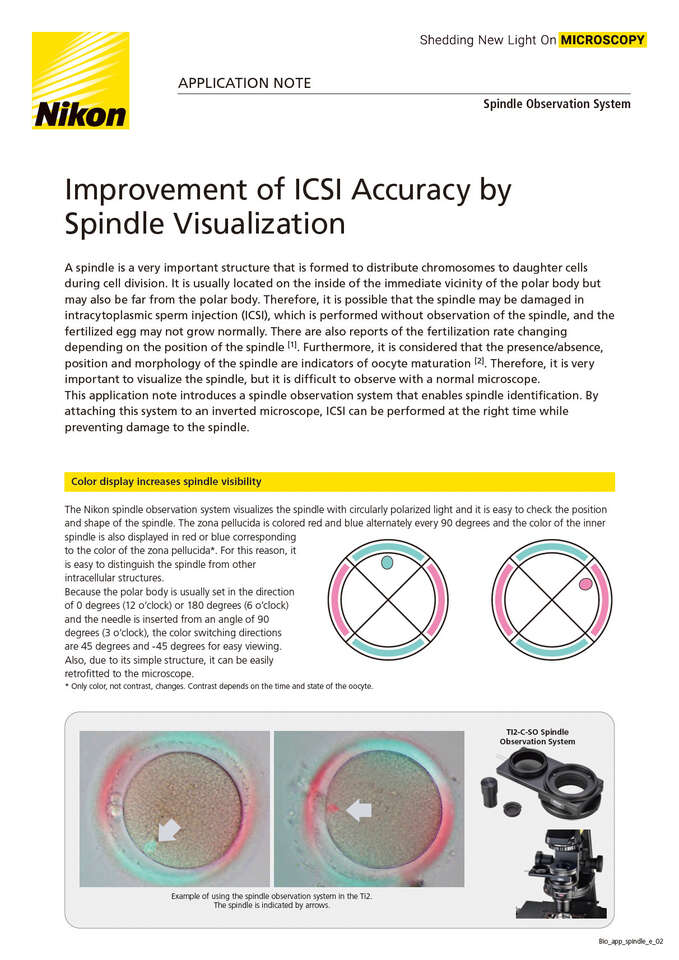
The Nikon spindle observation system visualizes the spindle with circularly polarized light and it is easy to check the position and shape of the spindle. The zona pellucida is colored red and blue alternately every 90 degrees and the color of the inner spindle is also displayed in red or blue corresponding to the color of the zona pellucida*. For this reason, it is easy to distinguish the spindle from other intracellular structures.
Because the polar body is usually set in the direction of 0 degrees (12 o’clock) or 180 degrees (6 o’clock) and the needle is inserted from an angle of 90 degrees (3 o’clock), the color switching directions are 45 degrees and -45 degrees for easy viewing.
Also, due to its simple structure, it can be easily retrofitted to the microscope.
* Only color, not contrast, changes. Contrast depends on the time and state of the oocyte.
Spindles are not always formed and it can be difficult to observe the spindle depending on the maturity of the oocyte. When the spindle cannot be confirmed even using this product, it is possible that the spindle is not formed or the spindle is not sufficiently mature. Normally, it is said that the spindle can be observed in the MII stage 6-8 hours after oocyte collection.
The spindle is very sensitive to temperature environment (around 37˚C is appropriate). When the temperature conditions are not appropriate, spindles cannot be observed due to the depolymerization of microtubules. It is recommended to use a “glass heater” for temperature environment management.
(Photo shows a product of Tokai Hit Co., Ltd.)
Use a glass-bottom container. Plastic-bottom containers cannot be used.
In the spindle observation system, a pair of the polarizing plate and the 1/4 wave plate is inserted into the optical path to irradiate the sample with circularly polarized light. Therefore, a certain amount of light is required to visualize the spindle.
Remove the ND filter from the fixed filter slot to secure the amount of light, increase the brightness of the light source and switch the optical path to 100% binocular port (in the case of using a binocular tube) instead of camera port.
Place one piece of the washer on the objective side module and attach the objective.
* Please note that the objective and the module will come into contact if the washer is not attached.
| NAMC observation → Spindle observation | Spindle observation → NAMC observation | |
|---|---|---|
| Condenser turret | Rotate to a “Blank” position | Place the NAMC condenser module into the optical path |
| Slider | Place the module for spindle observation (left side) into the optical path (Slide to the right) |
Place the NAMC/IMSI polarizer (right side) into the optical path (Slide to the left) |
Images courtesy of, Reproduction Clinic Tokyo
Relationship between pre-ICSI meiotic spindle angle, ovarian reserve, gonadotropin stimulation, and pregnancy outcomes.
J Assist Reprod Genet . 2017 May; 34(5): 609–615. Alina M. Mahfoudh, Jeong H. Moon, Sara Henderson, Elena Garcia-Cerrudo, Weon-Young Son, and Michael H. Dahan
Egg maturity assessment prior to ICSI prevents premature fertilization of late-maturing oocytes.
J Assist Reprod Genet . 2019 Mar; 36(3): 445–452. Zuzana Holubcová, Drahomíra Kyjovská, Martina Martonová, Darja Páralová, Tereza Klenková, Pavel Otevřel, Radka Štěpánová, Soňa Kloudová, and Aleš Hampl


