Nikon Europe B.V. | Europe & Africa
- en Change Region
- Global Site
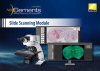
The new slide scanning solution from Nikon brings together the best of both worlds: a user friendly, yet extremely powerful software module that has been developed for robust, reliable hardware. Using the ECLIPSE Ni-E microscope in combination with the brand new NIS-Elements Slide Scanning module, finally there is no need to sacrifice image quality for the sake of simplicity.
This product is Research use only and should not be used for in vitro diagnostics.
This product is available in Europe. Please contact your local distributors or in-country representative for product availability in other regions.
Follow each step and use already predefined settings to get started with scanning.
No previous imaging experience needed.
*Fluorescence sample courtesy of NSALabs.com © All rights reserved.
To make your scanning workflow even smoother, our AI can automatically read sample labels, detect regions to be scanned, find the most optimal light settings, and ensure that your sample is always in focus. Low quality images can finally become a thing of the past.
The NIS-Elements Slide Scanning module supports both brightfield and fluorescence imaging, providing flexibility for diverse imaging applications.
Fluorescence staining of coronal section of mouse brain
Sample courtesy of NSALabs.com © All rights reserved.
Mouse embryo stained with hematoxylin eosin
Collecting data fast shouldn’t mean you have to compromise on image quality. The combination of Nikon’s renowned optics with the NIS-Elements Slide Scanning module allows you to get the best image quality in as little time as possible.
CFI Plan Apochromat Lambda D series objectives
Human lymph node stained with hematoxylin eosin
Acquiring your desired images shouldn’t take long. You can scan entire tissue sections even under a minute* making use of our fast stage and precise tiling.
* See specifications for details
Fluorescence staining of coronal section of mouse brain
Sample courtesy of NSALabs.com © All rights reserved.
Ensure accuracy by instantly comparing your scanned image side-by-side with a live sample at any time. With the synchronizer tool, navigating between the two is smooth and intuitive.
You can easily find and visualize any of your scanned images with the convenient gallery view.