![](https://downloads.microscope.healthcare.nikon.com/production/imager/mastheads/314/DS-Qi2-masthead-31_f076438097b2d2d85c6c3ef04742411e.jpg)
Capture low light fluorescence and large fields of view.
Equipped with a Nikon digital SLR camera FX-format CMOS sensor optimized for microscopy, the DS-Qi2 is an ultra-high quality 16.25 megapixel monochrome camera that features high pixel density, high sensitivity and low noise. The DS-Qi2 is an excellent choice for applications in quantitative fluorescence imaging.
* DS-Qi2 is not for clinical diagnostic use.
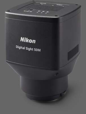
![](https://downloads.microscope.healthcare.nikon.com/production/imager/productphotos/313/DS-Qi2_2c4570626e41e228b41e37dfae7570ed.jpg 1x, https://downloads.microscope.healthcare.nikon.com/production/imager/productphotos/313/DS-Qi2_a8cc68e7194730b6cbe48ee0a564d9c9.jpg 2x)
Key Features
Large format CMOS sensors
Nikon manufactures CMOS sensors and imaging technologies for professional D-SLR cameras, which are now optimized for microscopy.
![](https://downloads.microscope.healthcare.nikon.com/production/imager/keyfeatures/Digital-Cameras/1502/DS-Ri2-CMOS-Sensors_eab673dc52b0446b2b598a80abc7b050.jpg 1x, https://downloads.microscope.healthcare.nikon.com/production/imager/keyfeatures/Digital-Cameras/1502/DS-Ri2-CMOS-Sensors_9b9989dbe1d056a5c35107959161a1c5.jpg 2x)
High sensitivity
Detects even faint fluorescent signals
7.3µm pixels, high quantum efficiency, and very low read noise allow the DS-Qi2 to even read faint fluorescent signals.
![](https://downloads.microscope.healthcare.nikon.com/production/imager/keyfeatures/Digital-Cameras/1514/DS-Qi2-signals_eab673dc52b0446b2b598a80abc7b050.jpg 1x, https://downloads.microscope.healthcare.nikon.com/production/imager/keyfeatures/Digital-Cameras/1514/DS-Qi2-signals_9b9989dbe1d056a5c35107959161a1c5.jpg 2x)
Pig kidney epithelial cells expressing GFP-EB3 tubulin
Sample courtesy of: Michael Davidson, National High Magnetic Field Laboratory, Florida State University
Excellent linearity
Reliable quantitative analysis made possible
With a linearity error of ±1%, the DS-Qi2 is a superb tool for measuring intensities in fluorescence samples, including time-based intensity measurement and ratiometric measurement.
Low noise
Acquires dim fluorescent signals with ultra-low noise
2.2 electrons read noise, coupled with a large full-well capacity, allows for the acquisition of fluorescence images with very little noise and a very high dynamic range.
![](https://downloads.microscope.healthcare.nikon.com/production/imager/keyfeatures/Digital-Cameras/1529/DS-Qi2-low-noise1_eab673dc52b0446b2b598a80abc7b050.jpg 1x, https://downloads.microscope.healthcare.nikon.com/production/imager/keyfeatures/Digital-Cameras/1529/DS-Qi2-low-noise1_9b9989dbe1d056a5c35107959161a1c5.jpg 2x)
![](https://downloads.microscope.healthcare.nikon.com/production/imager/keyfeatures/Digital-Cameras/1530/DS-Qi2-low-noise2_eab673dc52b0446b2b598a80abc7b050.jpg 1x, https://downloads.microscope.healthcare.nikon.com/production/imager/keyfeatures/Digital-Cameras/1530/DS-Qi2-low-noise2_9b9989dbe1d056a5c35107959161a1c5.jpg 2x)
LLC-PK1 cells expressing GFP-EB3 tubulin Low noise and large linear full well capacity allows the acquisition of large dynamic range in a single capture.
A captured image is shown intensity scaled to show both the brightest and dimmest areas.
Sample courtesy of: Michael Davidson, National High Magnetic Field Laboratory, Florida State University
Time-lapse photography
Fluorescent time-lapse imaging through integration with NIS-Elements software
With a large field of view, high pixel density, and low noise, the DS-Qi2 is ideal for time-resolved imaging applications.
![](https://downloads.microscope.healthcare.nikon.com/production/imager/keyfeatures/Digital-Cameras/1533/DS-Qi2-timelapse_eab673dc52b0446b2b598a80abc7b050.jpg 1x, https://downloads.microscope.healthcare.nikon.com/production/imager/keyfeatures/Digital-Cameras/1533/DS-Qi2-timelapse_9b9989dbe1d056a5c35107959161a1c5.jpg 2x)
![](https://downloads.microscope.healthcare.nikon.com/production/imager/keyfeatures/Digital-Cameras/1532/DS-Qi2-timelapse2_eab673dc52b0446b2b598a80abc7b050.jpg 1x, https://downloads.microscope.healthcare.nikon.com/production/imager/keyfeatures/Digital-Cameras/1532/DS-Qi2-timelapse2_9b9989dbe1d056a5c35107959161a1c5.jpg 2x)
Time-lapse images (every 1 second) of LLC-PK1 cells with GFP-EB3 tubulin. Each image represents the maximum intensity projection of the timelapse, allowing visualization of the end-binding protein located on the microtubule plus-ends, and allowing tracing of the microtubule path.
DS-Qi2 captures an extremely large field of view, but still represents very fine details as demonstrated in this cropped timelapse sequence from a large FOV image.
Objective: CFI Plan Apochromat Lambda 60X Oil (NA: 1.4)
Sample courtesy of: Michael Davidson, National High Magnetic Field Laboratory, Florida State University