Nikon Instruments Inc. | Americas
- en Change Region
- Global Site

Center of Excellence
The University Imaging Centers (UIC) serve internal and external research clients in the design of imaging experiments, choice of and training on suitable imaging systems, and subsequent image processing, visualization and analysis. The Centers provide a number of advanced imaging systems including: N-SIM super-resolution microscopy; macro spectral confocal microscopy; wide-field light and fluorescence microscopy; laser-scanning confocal microscopes; total internal reflectance microscope (TIRF); laser capture micro-dissection; several live cell imaging systems; poster printers; full sample preparation capabilities; along with 4 full-time and 4 part-time experienced staff members.
The UIC operates on the following fundamental mission goals:
email hidden; JavaScript is required
This A1si point scanning confocal system is configured on a Ti2-E inverted microscope and features a spectral detector for linear unmixing of signals from multiple emitters.
ECLIPSE Ti2-E inverted microscope with Perfect Focus System 4 (PFS4)
NIS-Elements software
This system features an A1R resonant scanning confocal system with Picoquant FLIM module for fluorescence lifetime imaging applications.
NIS-Elements software
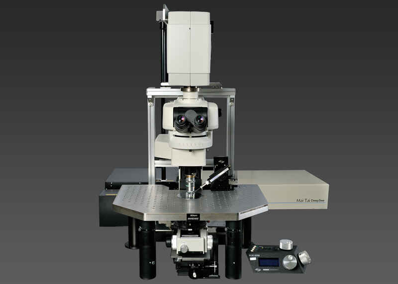
This A1R HD MP based resonant scanning system provides both fast multiphoton and visible imaging on one microscope.
ECLIPSE FN1 upright microscope
NIS-Elements software
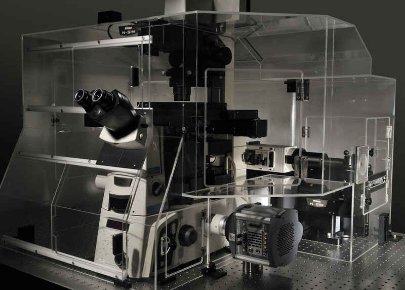
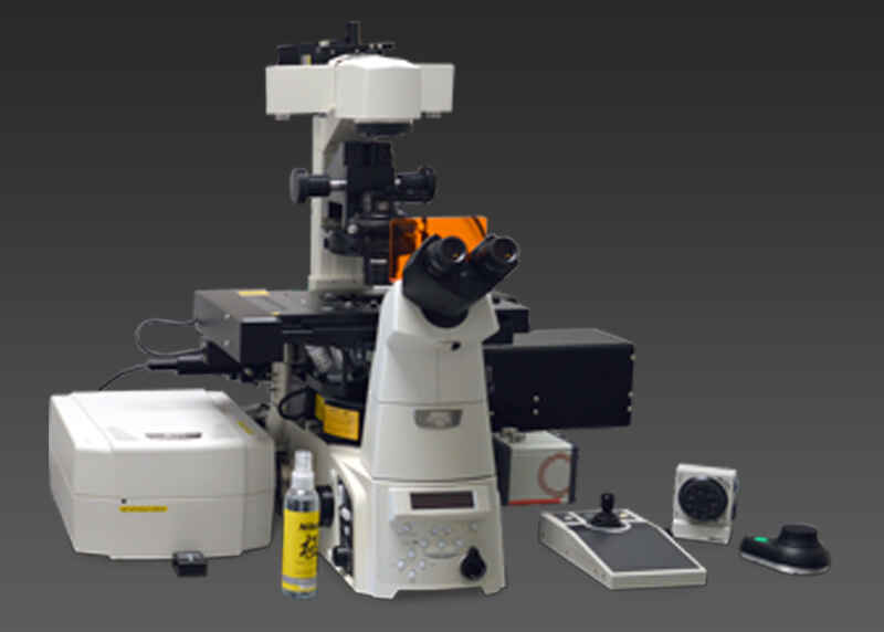
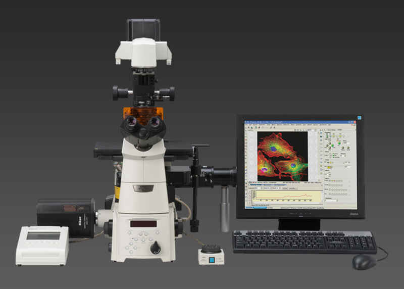
Fast confocal and super-resolution on the same Ti-E inverted microscope with the A1Rsi resonant scanning confocal and N-SIM structured illumination systems.
NIS-Elements software
This unique macro confocal system combines the AZ100 multizoom microscope with the C1si point-scanning confocal system with spectral detector.
NIS-Elements software
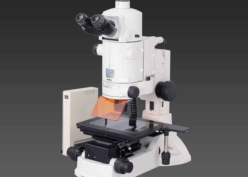
The AZ100M is the motorized version of the multizoom microscope/macroscope, and features multiple cameras and NIS-Elements software for recording observations.
NIS-Elements software
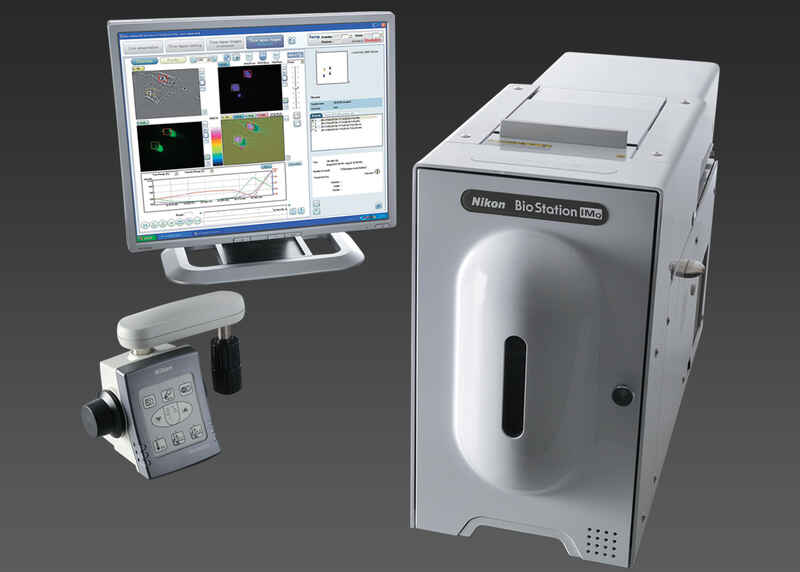
The BioStation IM-Q incorporates a microscope, an incubator and a high-sensitivity cooled CCD camera in a compact body. This all-in-one package provides a stable environment for live cells and advanced solutions for simple long-term time-lapse data acquisition.
The BioStation IM-Q incorporates a microscope, an incubator and a high-sensitivity cooled CCD camera in a compact body. This all-in-one package provides a stable environment for live cells and advanced solutions for simple long-term time-lapse data acquisition.
This widefield observation system is configured on a 90i upright microscope and features both monochrome sCMOS and color CCD cameras.
NIS-Elements software
This widefield observation system is configured on a TE2000-U invert4d microscope and features both monochrome and color CCD cameras.
NIS-Elements software
This system, based on the TE2000 inverted microscope features both sCMOS camera and NIS-Elements software, great for most routine applications.
NIS-Elements software
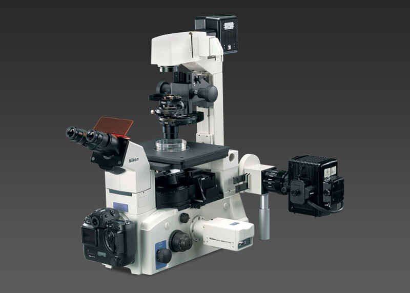
This live-cell imaging system is configured on a Ti-E inverted microscope and features incubation, deconvolution, and hardware triggering.
NIS-Elements software
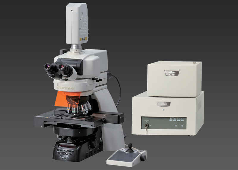
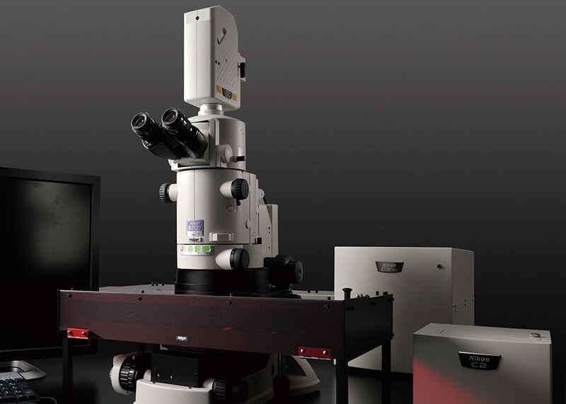
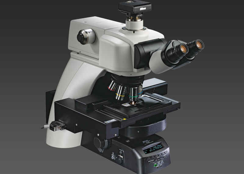
This C2 confocal system is configured on an Ni-E upright microscope and great for applications requiring optical sectioning.
ECLIPSE Ni-E upright microscope
C2 point scanning confocal system
NIS-Elements software
