- en Change Region
- Global Site
- Home
- Nikon BioImaging Centers
- Aix-Marseille University

Center of Excellence
Aix-Marseille University
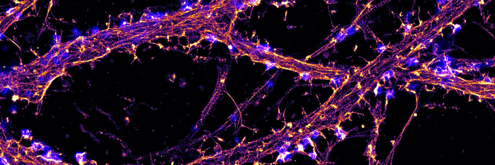
Nikon and the Neurophysiopathology Institute at Aix-Marseille University have partnered to create the Nikon Center of Excellence for Neuro-NanoImaging within its cellular imaging facility, focusing on how the latest super-resolutive technique can help in understanding brain cells and their dysfunctions.
The Center uniquely offers three different super-resolution microscopy techniques that are tailored for the subcellular investigation of neuronal function:
- Super-resolved spinning disk (Nikon SoRa) that provides fast, gentle imaging with optical sectioning at ~150 nm resolution, ideal for live-cell imaging.
- Structured Illumination Microscopy (Nikon N-SIM) that allows for robust, multicolor imaging with a 2x gain (~120 nm laterally) compared to diffraction-limited fluorescence microscopy.
- Stochastic Optical Reconstruction Microscopy (Nikon N-STORM) that reaches for ~20 nm detail to dissect the nanoscale organization of macromolecular complexes directly in cells.
The Nikon Center of Excellence at INP brings these cutting-edge techniques to all neurobiology researchers at INP, in Marseille and beyond, with expert advice on how to choose the best technique for a given question and project. New sample preparation, imaging procedures, and processing methods are developed based on the Center of Excellence microscopes within the facility.
Contact
Academic Director
email hidden; JavaScript is required
Facility Manager
email hidden; JavaScript is required
Address
Institute of NeuroPhysiopathology (INP)- UMR 7051
Faculty of Medical and Paramedical Sciences
27 Boulevard Jean Moulin
13385 Marseille Cedex 5, France
Systems Available

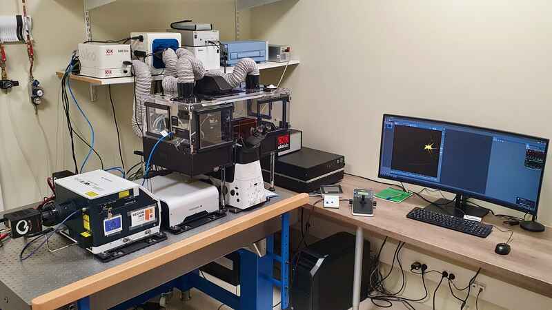
CSU-W1 SoRa Spinning Disk Confocal and Super-Resolution System
The CSU-W1 provides fast spinning disk confocal performance with large fields of view and deep in samples. Two cameras allow for simultaneous 2-color acquisition. In addition, the SoRa super-resolution module, based on optical pixel reassignment, can reach resolutions of 170 nm laterally, 350 nm axially with no compromise on the acquisition speed.
Components
ECLIPSE Ti2-E inverted microscope with Perfect Focus System 4 (PFS4)
- Yokogawa CSU-W1 spinning disk confocal with SoRa super-resolution module
- 2x Hamamatsu FusionBT sCMOS cameras
- Mad City Labs Nano-Z series Z-axis piezo stage
- OkoLab enclosure incubation system
NIS-Elements Advanced Research with 2D/3D Deconvolution and Clarify.ai

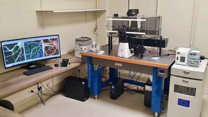
N-SIM Super-Resolution System
The N-SIM provides super-resolution Structured Illumination Microscopy (SIM) on the Ti2-E inverted microscope. It can reach a resolution of 120 nm laterally, 250 nm axially and is compatible with live-cell imaging.
Components
ECLIPSE Ti2-E inverted microscope with Perfect Focus System 4 (PFS4)
N-SIM S super-resolution
LUN-V 4-line laser unit (405 nm, 488 nm, 561 nm, 647 nm)
- Gemini image splitter and two Hamamatsu FusionBT sCMOS cameras
- Tokai Hit stage top incubator
- Mad City Labs Nano-Z series Z-axis piezo stage
- NIS-Elements Advanced Research with 2D/3D reconstruction and Enhance.ai

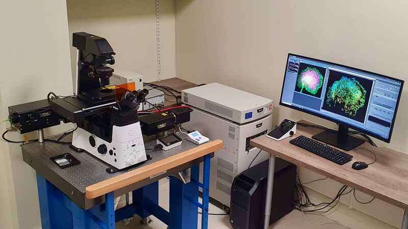
N-STORM/TIRF Super-Resolution System
The N-STORM provides super-resolution single molecule localization imaging on the Ti2-E inverted microscope. It can reach a resolution of 20 nm laterally, 50 nm axially, and also features live-cell TIRF imaging.
Components
ECLIPSE Ti2-E inverted microscope with Perfect Focus System 4 (PFS4)
- Motorized TIRF Illuminator Unit
LUN-V 4-line laser unit (405 nm, 488 nm, 561 nm, 647 nm)
- Hamamatsu FusionBT sCMOS camera
- Mad City Labs Nano-Z series Z-axis piezo stage
- Okolab stage-top incubation system
- NIS Elements software with N-STORM module
- Home
- Nikon BioImaging Centers
- Aix-Marseille University
