株式会社ニコンソリューションズ | Japan
- ja Change Region
- Global Site
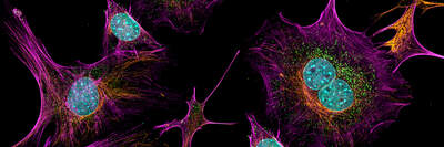
さまざまな2Dモデルシステムのイメージングサービスを提供します(幅広いアッセイのポートフォリオ、生細胞/固定細胞のスライド・ディッシュ・マルチウェルプレートサンプルのイメージング、落射蛍光顕微鏡や共焦点顕微鏡によるイメージング、インテリジェント画像取得及びAI解析など)。当ラボのアッセイポートフォリオは幅広い分野に対応しており、ユーザーのニーズに合わせた高度なカスタマイズが可能です。
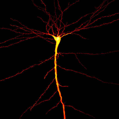
さまざまな3Dモデルシステムのイメージングサービスを提供します(幅広いアッセイのポートフォリオ、生細胞/固定細胞イメージング、タイムラプスイメージング、生物/組織全体のイメージング、落射蛍光顕微鏡や共焦点顕微鏡によるイメージング、インテリジェント画像取得及びAI解析など)。当ラボのアッセイポートフォリオは幅広い分野に対応しており、ユーザーのニーズに合わせた高度なカスタマイズが可能です。
Advanced spatial biology technologies to perform high-plex spatial imaging and whole transcriptome spatial profiling. Assay functional markers, protein biomarkers, chemokines, cytokines and RNA within tissues, all while retaining spatial context.
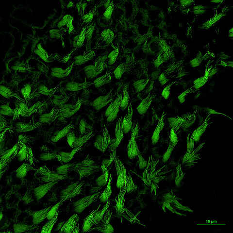
Unlock the power of ultra-detailed cellular imaging with super-resolution technology. Achieve unmatched clarity, ultra-low noise, and heightened sensitivity—ideal for revealing fine biological structures that standard microscopy can’t resolve.
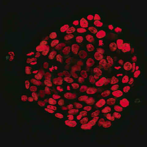
smFISH(single molecule fluorescence in situ hybridization)法は、単一のRNA分子を検出・定量する技術であり、単一細胞における遺伝子発現の時空間パターンの解析に幅広く使用されています。NBILでは、この技術を利用して、新薬候補化合物に対する遺伝子発現解析や空間生物学(spatial biology)的解析などのRNA定量サービスを提供しています。
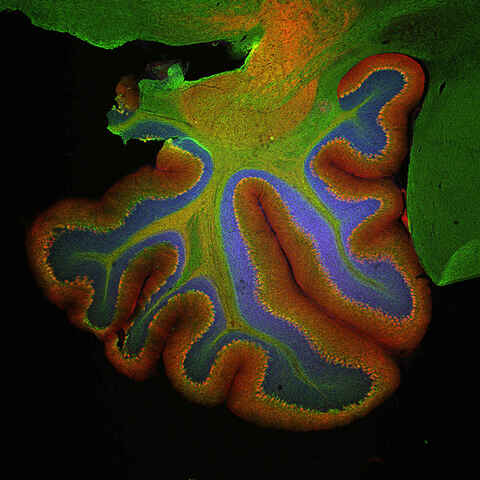
組織学、病理学、空間生物学(spatial biology)的解析、及び遺伝子発現解析のため、固定標本の明視野または蛍光によるスライド全体のスキャン/イメージングサービスを提供します。
Comprehensive imaging-based approaches for your toxicology studies during early drug development, drug safety assessments, and studies in MoA. Assess compound efficacy and efficiency in 2D and 3D cell culture models.
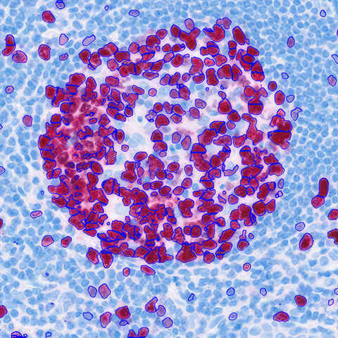
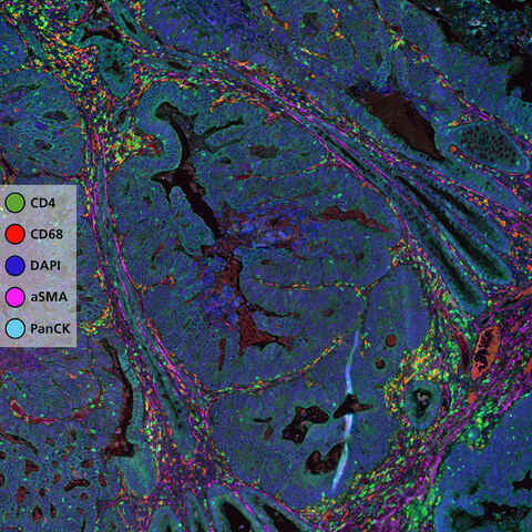
Customizable, automated, bias-free image acquisition and analysis for histology and pathology applications utilizing multiplexed chromogenic and fluorescent IHC, IF and ISH.
Generate consistent, high-quality data with Nikon’s turnkey automated microscopy and image analysis solutions.
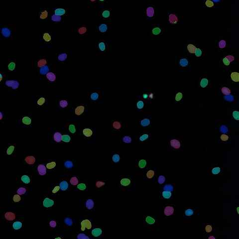
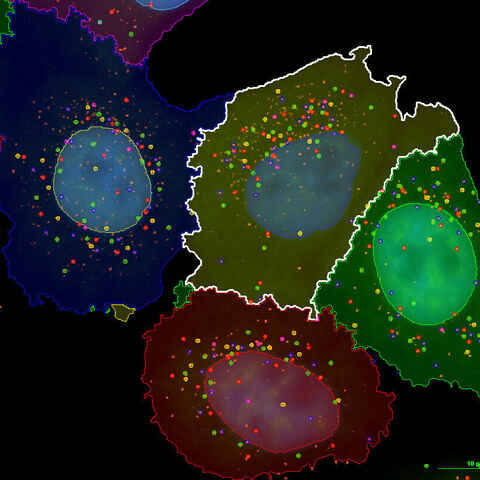
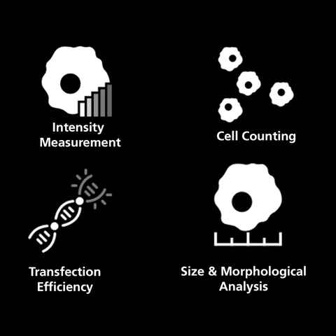
画像処理は、低シグナル画像の鮮明化から、安定的なセグメンテーションと定量化のための画像の作成まで幅広く活用されており、ほぼすべての顕微鏡実験で不可欠なプロセスとなります。カスタム解析ワークフローは、単一細胞データの収集から、ユーザーの定義する特徴を検出するためのAIネットワークのトレーニングまで、さまざまなアプリケーションに適応可能です。