- en Change Region
- Global Site
Application Notes

Resolving subcellular detail in tissue-mimetic culture
September 2024
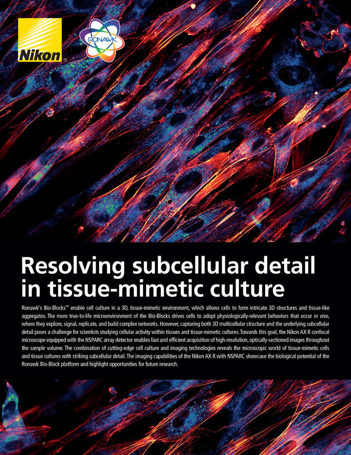
Ronawk’s Bio-Blocks™ enable cell culture in a 3D, tissue-mimetic environment, which allows cells to form intricate 3D structures and tissue-like aggregates. The more true-to-life microenvironment of the Bio-Blocks drives cells to adopt physiologically-relevant behaviors that occur in vivo, where they explore, signal, replicate, and build complex networks. However, capturing both 3D multicellular structure and the underlying subcellular detail poses a challenge for scientists studying cellular activity within tissues and tissue-mimetic cultures. Towards this goal, the Nikon AX R confocal microscope equipped with the NSPARC array detector enables fast and efficient acquisition of high-resolution, optically-sectioned images throughout the sample volume. The combination of cutting-edge cell culture and imaging technologies reveals the microscopic world of tissue-mimetic cells and tissue cultures with striking subcellular detail. The imaging capabilities of the Nikon AX R with NSPARC showcase the biological potential of the Ronawk Bio-Block platform and highlight opportunities for future research.
Bringing physiological context to cell studies
Research fields such as drug discovery and fundamental biology rely on experiments where cultured cells are studied in a variety of conditions. Many cell culture methodologies do not adequately mimic the complex 3D tissue environment in which cells naturally grow. This test system limitation fundamentally restricts what can be learned from cell culture experiments. Ronawk has developed a new cell culture platform called Bio-Blocks™ that mimic the physical properties of tissue. The macrostructure of the tissue-mimetic Bio-Block is a modular, 3D puzzle piece (Fig. 1A) that can interlock with other blocks (Fig.1B). The microstructure of the Bio-Block is a network of microchannels that facilitates cellular ingrowth in addition to permitting the perfusion of nutrients and gas exchange. Cells can proliferate across the surface of the block, in the microchannel network, and can even form dense, multicellular volumes at block-block interfaces (Fig. 1C). The tissue-mimetic environment of the Bio-Blocks beneficially impacts cell behavior and morphology, which can be observed via microscopy.
Visualizing 3D cellular organization and activity
Microscopy provides spatial context for intracellular, intercellular, and wider-scale multicellular structures, which is critical for understanding cellular health and activity. Widefield fluorescence microscopy is often unsuitable for imaging complex, 3D samples due to the nonspecific collection of out-of-focus light above and below the plane of interest (Fig. 1D, top left of diagonal divide). Instead, confocal microscopy utilizes a physical pinhole to reduce out-of-focus light and form clear images of cultured cells at a specific sample depth. This optical sectioning capability can be used to build volumetric reconstructions of various Bio-Block regions slice-by-slice in order to study the 3D structures formed by the cells. Utilizing the Nikon AX R confocal system equipped with the new NSPARC array detector and specialized Nikon objective lenses, we identify key tissue-mimetic behaviors and discern subtle details about cellular activity even when imaging 3D samples in situ. In this application note, we image adipose-derived stem cells cultured within the Bio-Block and stained to label nuclei, cytoskeletal structure, and both intra- and extracellular vesicles.
Specialized optics are critical for complex samples. As light passes through various media, its path is altered due to an interaction called refraction. Refraction is the basis for how a microscope objective lens collects light from a sample and projects it to form an image. However, variations in the refractive index (RI) of biological samples and cell culture substrates can introduce a variety of aberrations that limit the clarity of acquired images. Certain objective lenses are designed for use with specific immersion media (e.g., silicone oil) to better match the refractive index of the sample. Refractive index matching when conducting volumetric imaging is critically important. Closer matches reduce spherical aberration and minimize inaccuracies between the true sample and what is observed1. Improved image data quality enables more robust conclusions from each experiment. Utilizing Nikon’s diverse lineup of objective lenses, we conducted imaging with either water- or silicone-immersion (RI 1.33 and 1.41, respectively) lenses depending on whether the volume of interest contained predominately water-based media or cells, which have a higher RI than water (approximately 1.4).
Figure 1: Bio-Blocks enable 3D, tissue-mimetic cell culture. (A) Macro image of a Ronawk Bio-Block with central microchannel highlighted in red. (B) Diagram of interconnected Bio-Blocks and microchannel locations. (C) Interconnected Bio-Blocks imaged with a 2x Plan Apo λD objective lens on the AX R and zoomed to the approximate boxed region in panel B. Three regions of interest are highlighted: 1, where cells have proliferated into a block intrusion; 2, where cells have proliferated within a block-block interface; and 3, where cells have started to proliferate into one of the printed microchannels. (D) Comparison of widefield epifluorescence imaging (top left of diagonal line) versus confocal imaging (bottom right of diagonal line). (E) Cells in this application note were labeled with Hoechst 33342 to stain nuclei (405), Memglow™ 488 to stain the lipid membranes (488), phalloidin Texas Red to stain filamentous actin (F-actin) in the cytoskeleton (561), and wheat germ agglutinin Alexa Fluor™ 647 to stain glycoproteins in the plasma membrane (640). Cells were cultured for 14 days before fixation. Image acquired with the 40x Plan Apo λS silicone immersion objective and NSPARC array detector.
Enhanced image quality with NSPARC
Even with optimal optics, imaging relatively deep into large samples becomes difficult simply due to light attenuation. Depending on the features of interest, important details may become indistinguishable at depths of only tens to a few hundred micrometers within the sample. To maximize image quality, we used the new NSPARC detector, which features a 5 x 5 array of single pixel photon counting (SPPC) detectors in contrast to the single photomultiplier tube (PMT) detector used in traditional laser scanning confocal microscopy. This implementation of image scanning microscopy (ISM) provides a significant boost in both signal to noise ratio (SNR) and resolution2, which is extremely beneficial for imaging this complex sample.
Combining specialized objective lenses and NSPARC. We first compared the performance of an air objective paired with the Nikon DUX detector unit, a traditional PMT-based confocal detector, versus a water-immersion objective paired with the NSPARC detector to rapidly image a large sample volume at 20x magnification. The resonant scanning mode of the AX R confocal is well-suited for imaging relatively large volumes quickly, and plays a key role in improving the throughput of imaging experiments. Without proper refractive index matching, it is effectively impossible to distinguish subcellular details when imaging through 200 μm of water-based media between the dish bottom and the curved surface of a multicellular mass (Fig. 2). However, with proper refractive index matching and the enhanced image quality of the NSPARC detector, it becomes possible to visualize cellular details even when imaging at a fast rate (116 Z-sections with 1024 x 1024 pixel resolution (1024 px), covering a 442 x 442 x 204 μm volume in 73 seconds).
Better image quality enables higher imaging throughput. Capturing high-quality images can usually be achieved by increasing laser excitation power and exposure time at the cost of potential photodamage and slow scan speeds. We compared a DUX image optimized for quality (4096 px, galvo-scanning mode) to an NSPARC image optimized for acquisition speed (1024 px, resonant scanning mode), at a plane 75 μm deep into the curved multicellular mass (Fig. 3A). Qualitatively, the clarity of cellular features is similar between the two images, despite the galvo-scanned DUX image requiring 13 seconds per frame versus 0.5 seconds for the resonant-scanned NSPARC image. In order to image a 100 μm volume with this signal quality, field of view, and appropriate Z-step size, this translates to total acquisition times of 60 versus 5 minutes for the DUX and NSPARC cases, respectively. Sensitive samples and dyes degrade over time, so acquisition time can be critical. Of course, the 4k image has higher resolution potential due to the higher pixel density, but in practice this parameter can be tuned based on the size of the smallest feature to be resolved. These results demonstrate that imaging throughput can be substantially increased while maintaining resolution of subcellular features of interest by leveraging the elevated SNR offered by the NSPARC detector.
Figure 2: Optimized imaging conditions reveal cellular detail. (A) Depth-coded 3D view of cells proliferating off the edge of the block. The 442 x 442 x 204 μm volume was imaged in resonant scanning mode using the 20x water immersion objective and NSPARC detector. In total, 116, 1024 px frames were collected over 73 seconds. Solid grey line indicates underlying Bio-Block structure. (B, C) Comparison of cells imaged through 200 μm of media using (B) the 20x Plan Apo λD air objective and traditional confocal detector versus (c) the 20x Apo LWD λS water-immersion objective and NSPARC detector (C). This is a composite extended-depth of focus (EDF) image covering 5.28 μm of depth within the white-boxed region in panel A.
Figure 3: Maintaining image quality during fast acquisition with NSPARC. (A) Depth-coded 3D view of cells proliferating off the edge of the block. The 221 x 221 x 151 μm volume was imaged in resonant scanning mode using the 40x Plan Apo λS silicone immersion objective and NSPARC detector. In total, 151, 1024 px frames were collected over 76 seconds. The cutaway reveals the plane and region of interest for panels B and C. Solid grey line indicates underlying Bio-Block structure. (B, C) Comparison of cells imaged beneath 75 μm of combined media and cell mass using (B) the traditional confocal detector in galvo scanning mode with 4096 x 4096 pixel resolution versus (C) the NSPARC detector in resonant scanning mode with 1024 x 1024 pixel resolution.
Visualizing subcellular detail
We then performed a direct comparison of the DUX and NSPARC detectors combined with a silicone immersion objective and resonant scanning mode. We found that the NSPARC provided higher contrast and more clearly resolved subcellular details of cells proliferating 22 μm into a microchannel (Fig. 4).
Detecting cell-cell support behavior
The Bio-Blocks provide a structured, tissue-mimetic (micro)environment for cellular growth. In particular, one hallmark of multicellular tissue is mutual structural support between cells. We investigated the interface between Bio-Blocks to search for cells that are supported only by other cells, rather than the Bio-Block structure. Interestingly, we found a diversity of cell shapes and identified multiple instances of cells that were attached only to other cells (Fig. 5A-D). Similar behavior also occurs within the microchannels (Fig. 5E-H).
Single-cell level details indicate tissue-level behavior
The ability to resolve cytoskeletal features associated with endocytosis and exocytosis provides valuable insight into how cell culture systems can impact these processes. This visual information can include spatial arrangements of subcellular components and extracellular vesicles (EV). Ronawk has previously shown that the tissue-mimetic (micro)environment of their Bio-Blocks results in dramatically (7X) increased EV production relative to other cell culture platforms3. Using the NSPARC in combination with the optimization of imaging parameters, we maximized the resolving potential of the 40x silicone-immersion objective lens to visualize both the number of vesicles as well as the remarkable heterogeneity of the vesicular cargo itself (Fig. 6). For example, note the variety of 488 channel staining versus 640 channel staining of vesicles between adjacent cells. This diversity suggests variations in vesicular cargo composition or that the vesicles are produced via distinct mechanisms.
Figure 4: Direct comparison of DUX (single-point detector) and NSPARC (detector array). (A) Merged and channel views of a cell located 22 μm behind a mass of cells proliferating into a microchannel, imaged with the 40x Plan Apo λS silicone immersion objective and DUX detector in resonant scanning mode. 4.8 μm EDF. (B) The same location and imaging parameters recorded using the NSPARC detector.
Figure 5: Cell-cell support behavior fostered by the Bio-Block microenvironment. (A) Depth-coded 3D view of cells proliferating at the block-block interface. The 221 x 221 x 100 μm volume was imaged in resonant scanning mode using the 40x Plan Apo λS silicone immersion objective and NSPARC detector. In total, 201, 1024 px frames were collected over 120 seconds. The cutaway reveals plane of interest 18 μm beneath the surface cell layer. Solid gray lines indicate underlying Bio-Block structure. (B) Plane of interest featuring a variety of cell-cell contacts commonly seen in Bio-Block cell culture. 4 μm EDF. (C, D) Isolated nuclear and cytoskeletal channel views from boxed region in B. Arrowheads indicate points of cell-cell contact. (E) Depth-coded 3D view of cells proliferating at the block-block interface. The 204 x 204 x 32 μm volume was imaged in galvo-scanning mode using the 40x Plan Apo λS silicone immersion objective and NSPARC detector. In total, 39, 1024 px frames were collected over 40 seconds. The cutaway reveals plane of interest 10 μm beneath the surface cell layer. Solid gray circle indicates underlying microchannel structure. (F) Plane of interest featuring a variety of cell-cell contacts commonly seen in Bio-Block cell culture. 4 μm EDF. (G, H) Isolated nuclear and cytoskeletal channel views from boxed region in E. Arrowheads indicate points of cell-cell contact.
Figure 6: Diversity of cellular cargo revealed with NSPARC. (A) Full field of view image with 4096 x 4096 pixel resolution of surface level cells and underlying microchannel indicated by white circle. (B) Plane of interest 6 μm beneath the surface cell layer. Microchannel boundary indicated by white circle. Images acquired with the NSPARC detector and 40x Plan Apo λS silicone immersion in galvo-scanning mode. (C-E) Regions of interest indicated by white boxes in B with merged and single-channel views. Notable features include heterogenous vesicle staining patterns, different ratios of stained vesicles per cell, and subcellular localization of vesicles near the cytoskeleton or otherwise. White triangles indicate specific features of interest. Striped triangles indicate regions with more predominate staining in the 640 versus 488 nm channel.
Imaging complex, 3D samples in situ presents unique challenges. Nikon’s world-renowned objective lenses in combination with the new NSPARC array detector overcome these challenges, maximizing image quality and resolving subcellular details. With this optimized microscopy setup, we imaged cells cultured in Ronawk’s Bio-Blocks to study cell behavior and morphology. This allowed us to observe unique changes associated with cell culture in a tissue-mimetic environment. Combining this cutting-edge cell culture platform with advanced microscopy instrumentation offers a path toward more sophisticated experiments and advances in the fields of biologics production, bio-therapeutics, drug discovery, tissue engineering, and more.
References
- Diel, E.E., Lichtman, J.W. & Richardson, D.S. Tutorial: avoiding and correcting sample- induced spherical aberration artifacts in 3D fluorescence microscopy. Nat Protoc 15, 2773–2784 (2020). https://doi.org/10.1038/s41596-020-0360-2
- Colin J. R. Sheppard, Shalin B. Mehta, and Rainer Heintzmann, “Superresolution by image scanning microscopy using pixel reassignment,” Opt. Lett. 38, 2889-289w(2013). https://doi.org/10.1364/OL.38.002889
- Jacob G Hodge, Heather E. Decker, Jennifer L Robinson & Adam J Mellott. Tissue-mimetic culture enhances mesenchymal stem cell secretome capacity to improve regenerative activity of keratinocytes and fibroblasts in vitro. Wound Repair and Regeneration. February 2023; 31(3):367-383. https://doi.org/10.1111/wrr.13076
