Nikon Instruments Inc. | Americas
- en Change Region
- Global Site
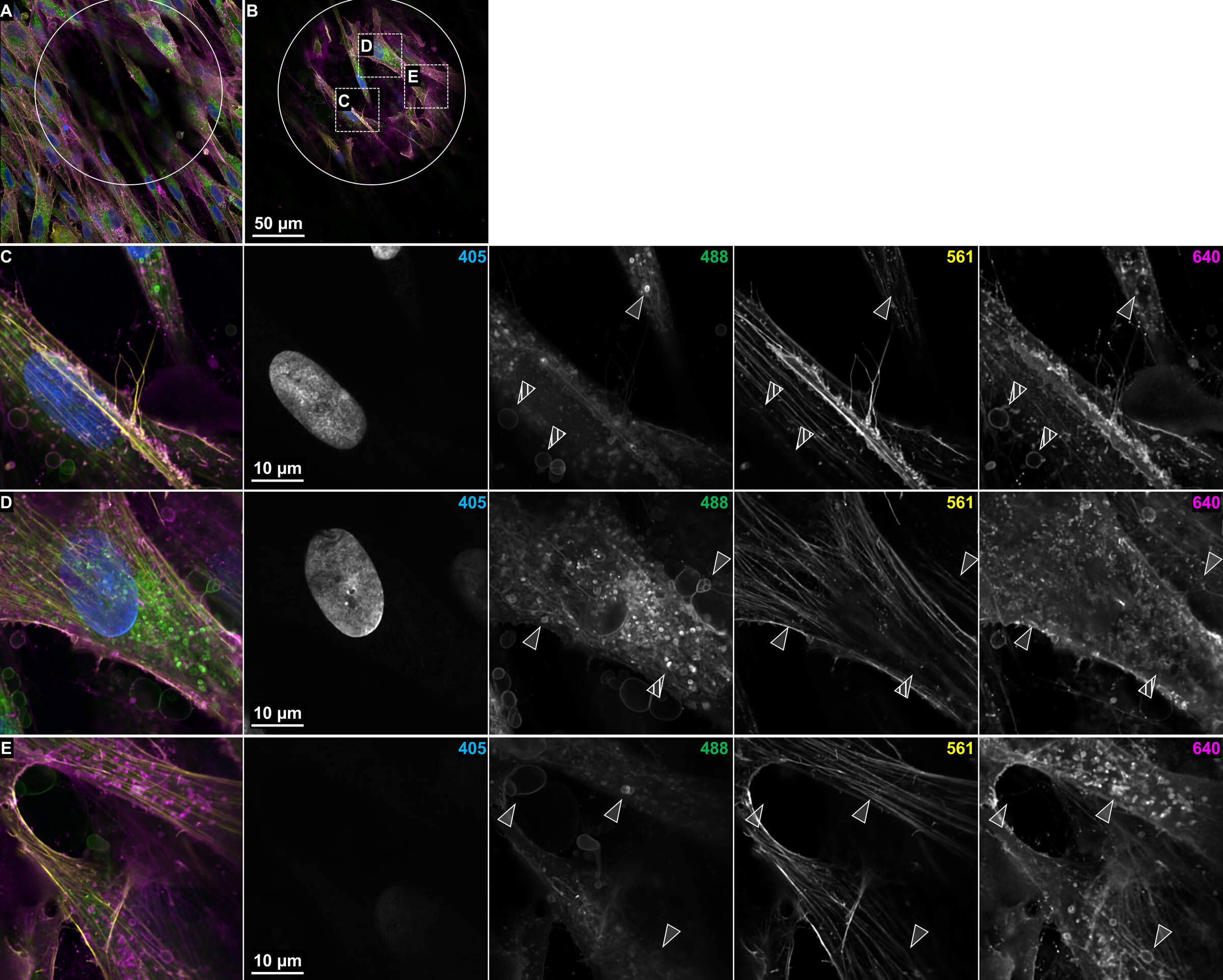
Figure 6: Diversity of cellular cargo revealed with NSPARC. (A) Full field of view image with 4096 x 4096 pixel resolution of surface level cells and underlying microchannel indicated by white circle. (B) Plane of interest 6 μm beneath the surface cell layer. Microchannel boundary indicated by white circle. Images acquired with the NSPARC detector and 40x Plan Apo λS silicone immersion in galvo-scanning mode. (C-E) Regions of interest indicated by white boxes in B with merged and single-channel views. Notable features include heterogenous vesicle staining patterns, different ratios of stained vesicles per cell, and subcellular localization of vesicles near the cytoskeleton or otherwise. White triangles indicate specific features of interest. Striped triangles indicate regions with more predominate staining in the 640 versus 488 nm channel.