Nikon Instruments Inc. | Americas
- en Change Region
- Global Site
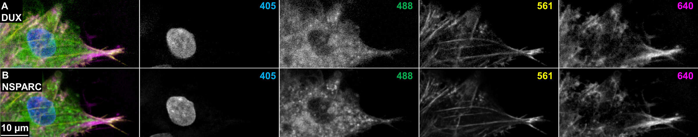
Figure 4: Direct comparison of DUX (single-point detector) and NSPARC (detector array). (A) Merged and channel views of a cell located 22 μm behind a mass of cells proliferating into a microchannel, imaged with the 40x Plan Apo λS silicone immersion objective and DUX detector in resonant scanning mode. 4.8 μm EDF. (B) The same location and imaging parameters recorded using the NSPARC detector.