Nikon Europe B.V. | Europe & Africa
- en Change Region
- Global Site
April 2024
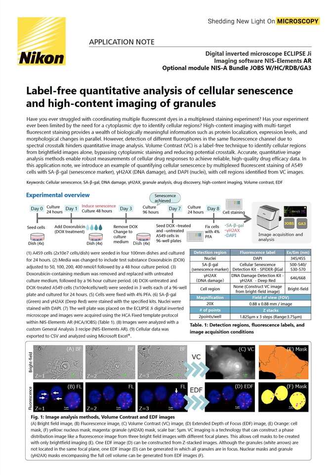
Have you ever struggled with coordinating multiple fluorescent dyes in a multiplexed staining experiment? Has your experiment ever been limited by the need for a cytoplasmic dye to identify cellular regions? High-content imaging with multi-target fluorescent staining provides a wealth of biologically meaningful information such as protein localization, expression levels, and morphological changes in parallel. However, detection of different fluorophores in the same fluorescence channel due to spectral crosstalk hinders quantitative image analysis. Volume Contrast (VC) is a label-free technique to identify cellular regions from brightfield images alone, bypassing cytoplasmic staining and reducing potential crosstalk. Accurate, quantitative image analysis methods enable robust measurements of cellular drug responses to achieve reliable, high-quality drug efficacy data. In this application note, we introduce an example of quantifying cellular senescence by multiplexed fluorescent staining of A549 cells with SA-β-gal (senescence marker), γH2AX (DNA damage), and DAPI (nuclei), with cell regions identified from VC images.