Nikon Europe B.V. | Europe & Africa
- en Change Region
- Global Site
April 2022
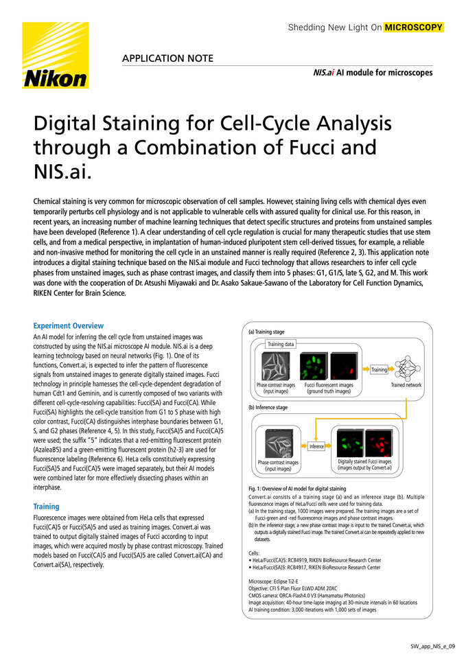
Chemical staining is very common for microscopic observation of cell samples. However, staining living cells with chemical dyes even temporarily perturbs cell physiology and is not applicable to vulnerable cells with assured quality for clinical use. For this reason, in recent years, an increasing number of machine learning techniques that detect specific structures and proteins from unstained samples have been developed (Reference 1). A clear understanding of cell cycle regulation is crucial for many therapeutic studies that use stem cells, and from a medical perspective, in implantation of human-induced pluripotent stem cell-derived tissues, for example, a reliable and non-invasive method for monitoring the cell cycle in an unstained manner is really required (Reference 2, 3). This application note introduces a digital staining technique based on the NIS.ai module and Fucci technology that allows researchers to infer cell cycle phases from unstained images, such as phase contrast images, and classify them into 5 phases: G1, G1/S, late S, G2, and M. This work was done with the cooperation of Dr. Atsushi Miyawaki and Dr. Asako Sakaue-Sawano of the Laboratory for Cell Function Dynamics, RIKEN Center for Brain Science.