Nikon Instruments Inc. | Americas
- en Change Region
- Global Site
August 2023
The light induced dimerization (LID) system, an optogenetic tool, uses photo-responsive proteins showing homo- or hetero-dimerization upon light illumination, allowing us to manipulate cell signaling.
This application note introduces an example of time-lapse imaging of activation and deactivation of the red/far-red responsive proteins expressed in HeLa cells, using the AX/AX R confocal microscope and NIR imaging option.
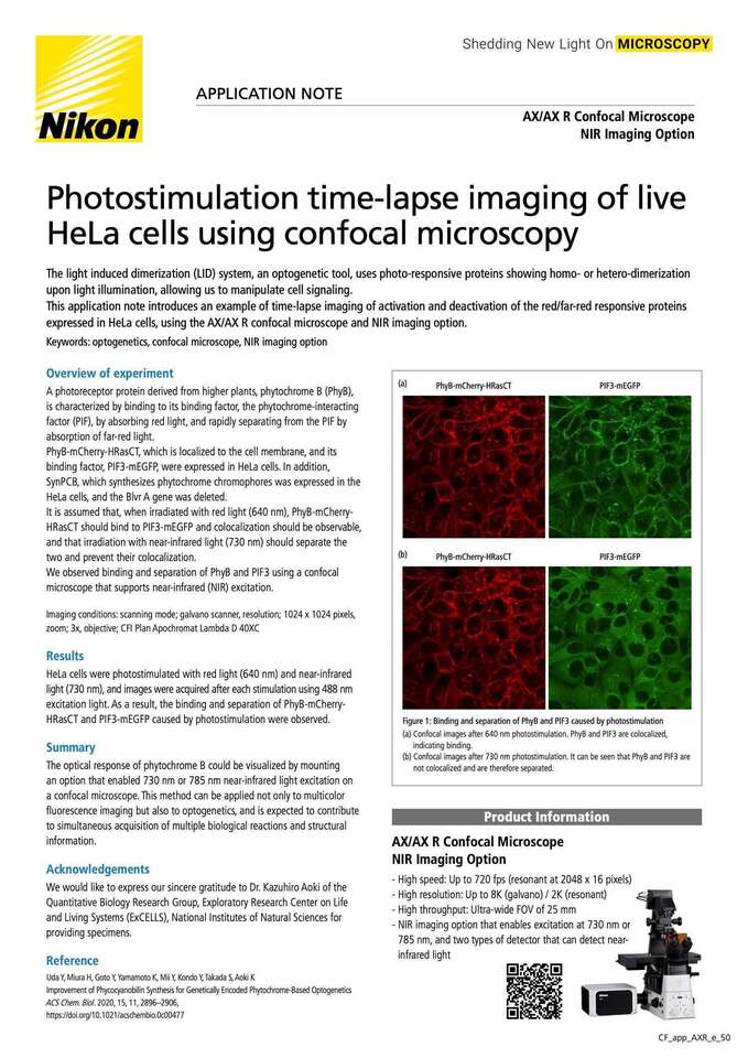
Figure 1: Binding and separation of PhyB and PIF3 caused by photostimulation
(a) Confocal images after 640 nm photostimulation. PhyB and PIF3 are colocalized, indicating binding.
(b) Confocal images after 730 nm photostimulation. It can be seen that PhyB and PIF3 are not colocalized and are therefore separated.
A photoreceptor protein derived from higher plants, phytochrome B (PhyB), is characterized by binding to its binding factor, the phytochrome-interacting factor (PIF), by absorbing red light, and rapidly separating from the PIF by absorption of far-red light.
PhyB-mCherry-HRasCT, which is localized to the cell membrane, and its binding factor, PIF3-mEGFP, were expressed in HeLa cells. In addition, SynPCB, which synthesizes phytochrome chromophores was expressed in the HeLa cells, and the Blvr A gene was deleted.
It is assumed that, when irradiated with red light (640 nm), PhyB-mCherry-HRasCT should bind to PIF3-mEGFP and colocalization should be observable, and that irradiation with near-infrared light (730 nm) should separate the two and prevent their colocalization.
We observed binding and separation of PhyB and PIF3 using a confocal microscope that supports near-infrared (NIR) excitation.
Imaging conditions: scanning mode; galvano scanner, resolution; 1024 x 1024 pixels, zoom; 3x, objective; CFI Plan Apochromat Lambda D 40XC
HeLa cells were photostimulated with red light (640 nm) and near-infrared light (730 nm), and images were acquired after each stimulation using 488 nm excitation light. As a result, the binding and separation of PhyB-mCherry-HRasCT and PIF3-mEGFP caused by photostimulation were observed.
The optical response of phytochrome B could be visualized by mounting an option that enabled 730 nm or 785 nm near-infrared light excitation on a confocal microscope. This method can be applied not only to multicolor fluorescence imaging but also to optogenetics, and is expected to contribute to simultaneous acquisition of multiple biological reactions and structural information.
We would like to express our sincere gratitude to Dr. Kazuhiro Aoki of the Quantitative Biology Research Group, Exploratory Research Center on Life and Living Systems (ExCELLS), National Institutes of Natural Sciences for providing specimens.
Uda Y, Miura H, Goto Y, Yamamoto K, Mii Y, Kondo Y, Takada S, Aoki KImprovement of Phycocyanobilin Synthesis for Genetically Encoded Phytochrome-Based OptogeneticsACS Chem. Biol . 2020, 15, 11, 2896–2906,
https://doi.org/10.1021/acschembio.0c00477
AX/AX R Confocal Microscope
NIR Imaging Option
These microscopes achieve high resolution images of 8K x
8K pixels, which is four times that of conventional models.
A large FOV with a diagonal of 25 mm allows acquisition
of a large area of samples in a single scan, reducing
phototoxicity. The AX R’s resonant scanner achieves a high
resolution of 2K x 2K pixels, allowing acquisition of live sample dynamics with
high-speed imaging of up to 720 fps
(2048 x 16 pixels).

