Nikon Instruments Inc. | Americas
- en Change Region
- Global Site
February 2026
Fluorescence microscopy is a widely-used approach for quantitatively analyzing the localization of a fluorescently-labeled target protein. However, fluorescence microscopy has a fundamental resolution limit that precludes true nanoscale resolution. In this application note, we introduce a study that addresses this limitation by employing Correlative Light-Electron Microscopy (CLEM), a technique that combines fluorescence and electron microscopy. Dr. Saeko Aoyama and Dr. Yusuke Hirabayashi from the Graduate School of Engineering, The University of Tokyo, applied a three-dimensional extension of CLEM, known as 3D CLEM, to uncover the molecular mechanisms underlying the formation of mitochondria–ER contact sites (MERCS) in mammalian cells. For fluorescence imaging, they used the AX with NSPARC super-resolution microscope system to capture high-resolution localization of MERCS regulatory molecules. Subsequently, they demonstrated that these localizations correspond to the fine structural details of MERCS using 3D CLEM1. This revealed that complexes formed by mitochondrial and ER-associated molecules play an essential role in MERCS formation.
1 CLEM was conducted in collaboration with Drs. Noboru Mizushima, Chieko Saito, Ikuko Honda, Akira Takahashi and Yoko Ishida from the Graduate School of Medicine, The University of Tokyo.
Keywords: Mitochondria, Endoplasmic Reticulum, MERCS, CLEM Method, AX with NSPARC
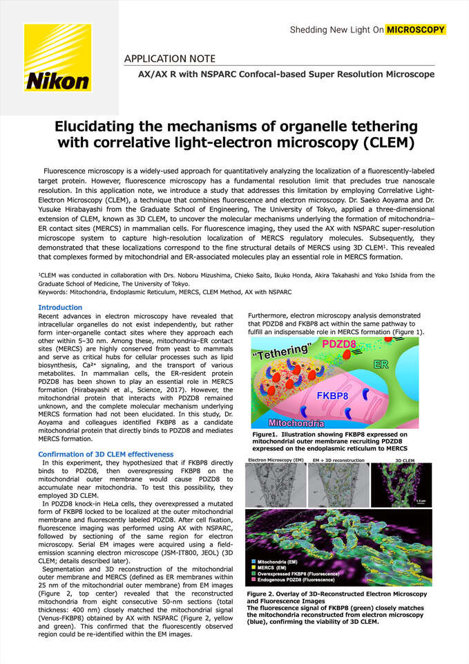
Figure1. Illustration showing FKBP8 expressed on mitochondrial outer membrane recruiting PDZD8 expressed on the endoplasmic reticulum to MERCS
Recent advances in electron microscopy have revealed that intracellular organelles do not exist independently, but rather form inter-organelle contact sites where they approach each other within 5–30 nm. Among these, mitochondria–ER contact sites (MERCS) are highly conserved from yeast to mammals and serve as critical hubs for cellular processes such as lipid biosynthesis, Ca²⁺ signaling, and the transport of various metabolites. In mammalian cells, the ER-resident protein PDZD8 has been shown to play an essential role in MERCS formation (Hirabayashi et al., Science, 2017). However, the mitochondrial protein that interacts with PDZD8 remained unknown, and the complete molecular mechanism underlying MERCS formation had not been elucidated. In this study, Dr. Aoyama and colleagues identified FKBP8 as a candidate mitochondrial protein that directly binds to PDZD8 and mediates MERCS formation.
Furthermore, electron microscopy analysis demonstrated that PDZD8 and FKBP8 act within the same pathway to fulfill an indispensable role in MERCS formation (Figure 1).
Figure 2. Overlay of 3D-Reconstructed Electron Microscopy and Fluorescence Images
The fluorescence signal of FKBP8 (green) closely matches the mitochondria reconstructed from electron microscopy (blue), confirming the viability of 3D CLEM.
In this experiment, they hypothesized that if FKBP8 directly binds to PDZD8, then overexpressing FKBP8 on the mitochondrial outer membrane would cause PDZD8 to accumulate near mitochondria. To test this possibility, they employed 3D CLEM.
In PDZD8 knock-in HeLa cells, they overexpressed a mutated form of FKBP8 locked to be localized at the outer mitochondrial membrane and fluorescently labeled PDZD8. After cell fixation, fluorescence imaging was performed using AX with NSPARC, followed by sectioning of the same region for electron microscopy. Serial EM images were acquired using a field- emission scanning electron microscope (JSM-IT800, JEOL) (3D CLEM; details described later).
Segmentation and 3D reconstruction of the mitochondrial outer membrane and MERCS (defined as ER membranes within 25 nm of the mitochondrial outer membrane) from EM images (Figure 2, top center) revealed that the reconstructed mitochondria from eight consecutive 50-nm sections (total thickness: 400 nm) closely matched the mitochondrial signal (Venus-FKBP8) obtained by AX with NSPARC (Figure 2, yellow and green). This confirmed that the fluorescently observed region could be re-identified within the EM images.
Strikingly, the fluorescence signal of endogenous PDZD8 was found accumulating at MERCS, as revealed by electron microscopy (arrowheads in Figure 3). Furthermore, quantitative analysis of endogenous PDZD8 overlapping with mitochondria in fluorescence images acquired using AX with NSPARC showed a significant increase upon FKBP8 overexpression (Figure 4).
Taken together, these findings suggest that overexpression of FKBP8 on the mitochondrial outer membrane increases the abundance of PDZD8–FKBP8 protein complexes at MERCS. Combined with other results, this demonstrates the essential role played by the PDZD8–FKBP8 complex in bridging the ER membrane and the mitochondrial outer membrane to form MERCS.
Figure 3. Overlay of Z projection of 3D electron microscopy reconstruction and fluorescence image
Endogenous PDZD8 was observed recruiting at MERCS (arrowheads).
Figure 4. The percentage of endogenous PDZD8 overlapping with mitochondria significantly increased upon FKBP8 overexpression.
Figure 5. Workflow of 3D CLEM
Fluorescence microscopy allows observation of fluorescently-labeled protein localization, but the resolution is insufficient for direct visualization of nanoscale structures such as MERCS. Therefore, using CLEM to combine the advantages of light and electron microscopy is a highly effective approach for clarifying protein localization at MERCS. In this study, PDZD8 was first observed by fluorescence microscopy, and then the same field was examined by electron microscopy to identify the position of MERCS. Specifically, AX with NSPARC was used to acquire high-resolution fluorescence images, and the corresponding regions were imaged by three-dimensional electron microscopy and correlated using 3D CLEM (Figure 5). When performing 3D CLEM, if the resolution gap between electron microscopy and fluorescence microscopy is too large, it becomes difficult to cover all optical sections of the fluorescence image in the EM dataset and to accurately align the two image sets. The super- resolution capability of AX with NSPARC produces higher resolution fluorescence images compared to conventional confocal microscopes, enabling overlay with EM images using a minimal number of serial sections.
This approach allowed three-dimensional visualization of PDZD8-derived fluorescence signals overlapping with the fine structure of MERCS.
In this study, by combining high-resolution PDZD8 localization analysis using AX with NSPARC and structural analysis of MERCS by electron microscopy, it was revealed that the mitochondrial outer membrane protein FKBP8 recruits the ER protein PDZD8 and regulates MERCS formation. These findings advance our understanding of cellular homeostasis, in which MERCS play a critical role.
Mutations in MERCS-related genes, including PDZD8, have been reported to be associated with several neurological disorders. A deeper understanding of MERCS dynamics is expected to lead to broad applications, from basic research to clinical and therapeutic approaches.
Nakamura K, Aoyama-Ishiwatari S, Nagao T, Paaran M, Obara CJ, Sakurai-Saito Y, Johnston J, Du Y, Suga S, Tsuboi M, Nakakido M, Tsumoto K, Kishi Y, Gotoh Y, Kwak C, Rhee HW, Seo JK, Kosako H, Potter C, Carragher B, Lippincott-Schwartz J, Polleux F, Hirabayashi Y. Mitochondrial complexity is regulated at ER- mitochondria contact sites via PDZD8-FKBP8 tethering. Nat Commun. 2025 Apr 17;16(1):3401. doi: 10.1038/s41467-025-58538-3.
We would like to express our sincere gratitude to Dr. Saeko Aoyama of the Graduate School of Engineering, The University of Tokyo, for kindly providing images and sharing valuable insights into the research for the preparation of this application note.
Edited by: Masaki Tatara, Nikon Corporation
AX/AX R with NSPARC Confocal-based Super Resolution Microscope
The super-resolution detector NSPARC, which features a 25-detector array, achieves even higher resolution with excellent S/N ratio, without impairing the functionality of the conventional AX/AX R confocal microscope.


