 ®
Nikon Instruments Inc. | Americas
- en Change Region
- Global Site
May 2026
Lipid nanoparticles (LNP) have progressed rapidly from experimental carriers of nanomedicines to clinically validated drug products. Despite this success, development and manufacturing workflows still rely on measurements that collapse complex nanoparticle populations into single average values. A single LNP formulation often contains a mixture of particles that differ in size, internal structure, lipid organization, and nucleic acid content. Some particles carry multiple mRNA molecules, others only one, and some none. These subpopulations can coexist within the same batch yet exhibit distinct biological behaviors that influence delivery efficiency, stability, immunotoxicity, and therapeutic response.
Standard analytical tools—including dynamic light scattering (DLS), nanoparticle tracking analysis (NTA), and bulk fluorescence assays—are not designed to resolve this diversity. They report ensemble averages that obscure particle-to-particle variability and provide little insight into how formulation parameters shape internal organization. As a result, formulation optimization, scale-up, and comparability assessments remain largely empirical, limiting the ability to rationally engineer next-generation nanomedicine therapeutics.
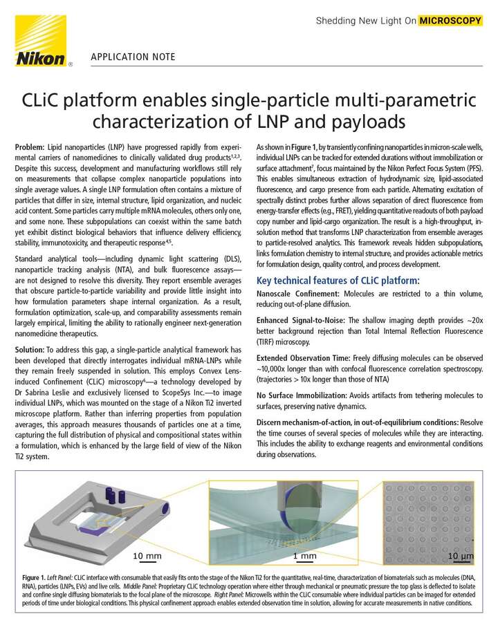
To address this gap, a single-particle analytical framework has been developed that directly interrogates individual mRNA-LNPs while they remain freely suspended in solution. This employs Convex Lens-induced Confinement (CLiC) microscopy6—a technology developed by Dr Sabrina Leslie and exclusively licensed to ScopeSys Inc.—to image individual LNPs, which was mounted on the stage of a Nikon Ti2 inverted microscope platform. Rather than inferring properties from population averages, this approach measures thousands of particles one at a time, capturing the full distribution of physical and compositional states within a formulation, which is enhanced by the large field of view of the Nikon Ti2 system.