Nikon Instruments Inc. | Americas
- en Change Region
- Global Site
March 2025
Microphysiological Systems (MPS) are 3D culture systems designed to mimic in vivo microenvironments. They are gaining attention as tools for preclinical pharmaceutical evaluation, offering a more physiologically-relevant human model. They also offer an ethical alternative to animal testing. MPS utilizes components such as microfluidic channels and porous membranes to faithfully recreate the microenvironment surrounding cells in 3D. This enables research in conditions that are closer to living tissues than those possible with conventional 2D cultures. MPS is already being applied in diverse research areas, including regenerative medicine, drug development, toxicity evaluation, and disease modeling, and is expected to become a new standard for in vitro evaluation.
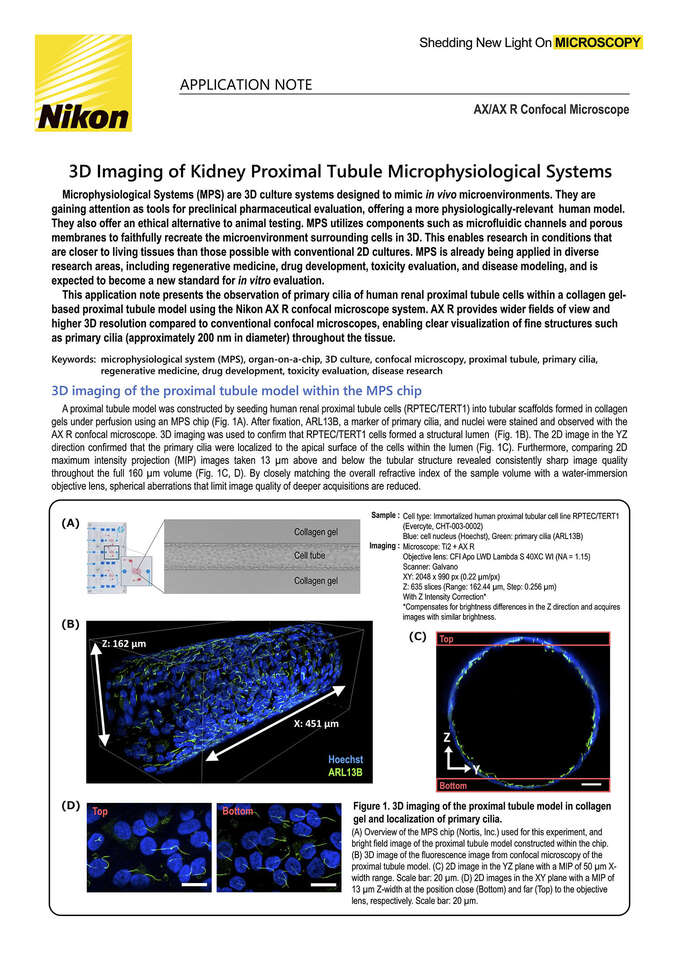
This application note presents the observation of primary cilia of human renal proximal tubule cells within a collagen gel-based proximal tubule model using the Nikon AX R confocal microscope system. AX R provides wider fields of view and higher 3D resolution compared to conventional confocal microscopes, enabling clear visualization of fine structures such as primary cilia (approximately 200 nm in diameter) throughout the tissue.
Keywords: microphysiological system (MPS), organ-on-a-chip, 3D culture, confocal microscopy, proximal tubule, primary cilia, regenerative medicine, drug development, toxicity evaluation, disease research