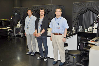
Tomomi Nemoto, Ph.D. and Ryosuke Kawakami, Ph.D.
Research Institute for Electronic Science
Sapporo, Hokkaido, Japan
Nikon Equipment

Please tell us about your research.
Based on the development of new imaging technologies and the discovery of mechanisms of the body using these technologies, especially the functions of organs, we conduct research in order to understand vital functions in hierarchical structures, such as molecules, cells and organs. In particular, we study brain neuroscience and exo- and endocrinology from a clinical perspective and the neuropathological aspects of neuroscience. We hope our research will contribute to the health and welfare of the public.
In the past, imaging technologies have been developed through the ingenuity of researchers, who discovered effective imaging conditions during their experiments. Continuous feedback between academics working in life sciences and on technological developments is very important. Thus, I believe understanding the real needs of researchers will lead to the development of more useful instruments.
In our laboratory, through focusing on high-quality deep imaging of living organisms, we study a wide range of vital phenomenon and different areas of the body. This also requires various fluorescence imaging techniques. Likewise, once a microscope is developed here that can visualize exocytotic events, it will be used also by outside collaborators to research insulin. In this way, our laboratory performs the role of a hub for multi-directional research.
What applications do you use the Nikon confocal microscope for?
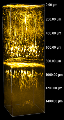
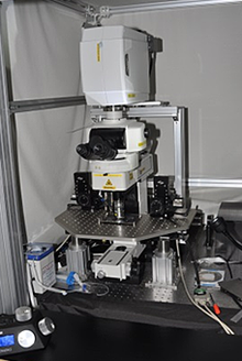
We use Nikon’s Multi-photon Confocal Microscope A1R MP as a two-photon microscope is effective for deep imaging of living organisms. We image molecular dynamics in the brain, skin, pancreas and adipose cells, using live mice and cell lines. In addition to the morphological change of minute cell structures, we observe the activities of various signal molecules in cells by use of fluorescence proteins and synthetic organic dyes. We do simultaneous imaging of both blood flow and morphological change of neurons in deep areas of the brain. Nowadays, we are beginning to utilize high order harmonic generation to visualize non-stained fibriform structures. We hope to expand its applications.
Why do you choose Nikon products?
Nikon’s multi-photon microscopes have sophisticated technologies. The quality of Nikon’s objective lenses and the design of Nikon’s Non Descanned Detector are excellent. They are very important for in vivo imaging, where Nikon is ahead of other manufacturers. We are impressed with the highly detailed images acquired by them. Also, we have only high praise for the Confocal Microscope A1 series and the high-resolution, high-quality images it produces.
Furthermore, we greatly appreciate Nikon’s technical support. We get meticulous answers to our highly technical questions.
What are the future demands for microscopes and imaging?
Firstly, we expect further improvements to basic performance, including spatial and temporal resolutions, as well as observation depth.
Secondly, I hope that spectral detectors can be more generally used. This is because I believe that with multi-color imaging even those not familiar with microscope operations should be able to acquire scientifically correct images based on accurate wavelength information. Confocal detectors using fluorescence filters can only acquire specific wavelength information. Therefore, the skill of selecting suitable filters is required to acquire reliable images. Unfortunately, incorrect interpretations of multi-staining images are sometimes seen in science literature.
Furthermore, I hope to get more information by effectively applying various non-linear optical phenomena. For example, I’d like to see microscopes that can extract image information from non-stained samples. These microscopes could be used for clinical applications in the future. As with multi-photon microscopes, which are now widely used, I believe non-linear optical systems would also be rapidly accepted if there are breakthrough innovations.
What are the future demands for Nikon?
I expect Nikon to become a world leader. Nikon products are extremely reliable and Nikon has developed remarkable technologies. I hope the company will accelerate the speed of its technical innovations. Nikon’s NIS-Elements software has the advantage of being compatible with different types of microscopes with common usability, I hope Nikon keeps improving in this direction.
And I also hope that researchers will be able to build their observation systems using Nikon microscopes more flexibly, in a way that will meet their own research needs. For example, in electrophysiology and behavioral analysis experiments, it is best if a whole experiment system, including microscopes and peripheral devices used, is under the integrated control of a single I/O board.
Please tell us about the Nikon Imaging Center at Hokkaido University
We think the aim of the Nikon Imaging Center at Hokkaido University (NIC@HU) to provide researchers with the opportunity to use current imaging equipment from Nikon and other manufacturers has been an overwhelming success. The number of people using NIC@HU has been steadily increasing thanks to our technical seminars and continuous support from businesses working in cooperation with the imaging center. We are proud to contribute to the education of young researchers not just at Hokkaido University but in Hokkaido Prefecture as a whole. In addition to the research needs of the university, experimental research commissioned by private businesses has been increasing. So too has the number of visitors from overseas interested in Japanese cutting-edge technologies. Requests for commissioned experimental research have come from outside Japan as well.
We believe it is important for the future to have a wide range of the latest imaging instruments at NIC@HU. Also, we hope to establish a mechanism through which we can provide feedback from our cutting-edge cooperative research with Nikon to general users.