- fr Change Region
- Global Site
Select Your Region and Language
Products and Promotions may differ based on your selected Region.
Americas
Europe & Africa
- Accueil
- Produits
- Microscopes Digitaux
- PreciPoint Microscope and Slide Scanners
PreciPoint Microscope and Slide Scanners
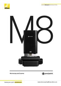
Dual digital microscope & slide scanner
Specifications
Microscope and Scanner
| M8 | O8 | |
|---|---|---|
Operating modes tailored to diverse workflows | Live-mode, InstantScan mode, SlideScan-mode | |
Light | Transmitted Light; LED, brightfield | |
Barcode scanning | Upon request | |
Supported objectives | 20x to 60x air | 20x to 100x air and oil |
Dimensions | 45 cm x 40 cm x 30 cm; 25 kg | |
Automated Microscope | X-Y-stage automated, Z-axis automated | |
Handling | Controlled with computer; Live remote control due to automation | |
Digitization
| M8 | O8 | |
|---|---|---|
Scanning parameters | Whole Slide Imaging or partial digitization | |
Scanning algorithms | Different scanning algorithms tailored to different slide qualities | |
InstantScan-mode | Large field of view within seconds at high resolution |
|
Scanning speed per slide | 20x: 2 min | 100x: 1 hr |
Scanning resolution per slide | 60x: 0.85 NA: 0.16 μm/px | 100x: 1.3 NA: 0.096 μm/px |
Slide capacity | 25 x 75 mm (2 slides) or 50 x 75 mm (1 slide) | |
Z-Axis Resolution | 25 nm | |
XY-Axis Resolution | 78 nm | |
Z-Stacking | Yes | |
Cloud, Image Analysis, and Computer
| M8 | O8 | |
|---|---|---|
Operating software | MicroPoint included | |
Viewer software | ViewPoint* included (unlimited users) | |
Storage | PreciCloud slide storage | |
Image analysis | Several software applications on request, based on customer needs | |
Computers | Various computers recommended and approved by PreciPoint | |
Microscope computer connection | USB 3.0 | |
Image output formats | PNG, JPEG, TIFF, BMP, VMIC, XLS, and many more | |
Microscopes Digitaux
- ECLIPSE Ji
- ECLIPSE Ui
- PreciPoint Microscope and Slide Scanners
Produits connexes
- Accueil
- Produits
- Microscopes Digitaux
- PreciPoint Microscope and Slide Scanners

