Nikon Europe B.V. | Europe & Africa
- en Change Region
- Global Site
November 2023
A picture is worth a thousand words
Since the time of Linnaeus, approximately 1.5 million new species have been described. As this accounts for only a modest fraction of global species diversity, taxonomists are constantly looking for methods to accelerate the pace of species discovery and enhance conventional description methods. Taxonomic descriptions depend on illustrations more than any other disciplines. From the earliest taxonomic treatments, the descriptions of species have nearly always been accompanied by visual representations to convey information about the morphology of the studied taxa. The importance of digital imaging in taxonomy has been long recognized, and its role in enhancing species description universally recognized.
Dr. Nesrine Akkari
Curator of the collection Myriapoda
Natural History Museum Vienna
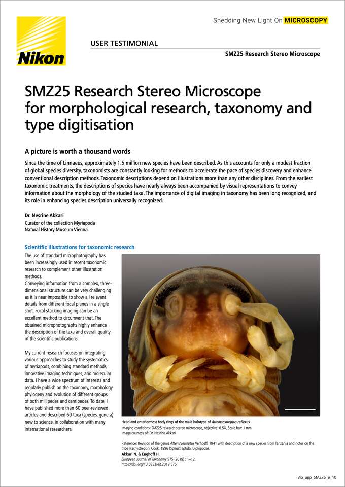
Head and anteriormost body rings of the male holotype of Attemsostreptus reflexus
Imaging conditions: SMZ25 research stereo microscope, objective: 0.5X, Scale bar: 1 mm
Image courtesy of: Dr. Nesrine Akkari
Reference: Revision of the genus Attemsostreptus Verhoeff, 1941 with description of a new species from Tanzania and notes on the tribe Trachystreptini Cook, 1896 (Spirostreptida, Diplopoda).
Akkari N. & Enghoff H.
European Journal of Taxonomy 575 (2019) : 1–12.
https://doi.org/10.5852/ejt.2019.575
The use of standard microphotography has been increasingly used in recent taxonomic research to complement other illustration methods.
Conveying information from a complex, three-dimensional structure can be very challenging as it is near impossible to show all relevant details from different focal planes in a single shot. Focal stacking imaging can be an excellent method to circumvent that. The obtained microphotographs highly enhance the description of the taxa and overall quality of the scientific publications.
My current research focuses on integrating various approaches to study the systematics of myriapods, combining standard methods, innovative imaging techniques, and molecular data. I have a wide spectrum of interests and regularly publish on the taxonomy, morphology, phylogeny and evolution of different groups of both millipedes and centipedes. To date, I have published more than 60 peer-reviewed articles and described 60 taxa (species, genera) new to science, in collaboration with many international researchers.
The Nikon SMZ25 research stereo microscope is routinely used to examine and illustrate key morphological details of the animal group I study. Equipped with a DS-Ri2 camera, it gives a high performance allowing me to examine and photograph external morphological details of the studied species, ranging from coarse features to minute structures. For my taxonomic work, as well as the ability to describe new taxa and prepare scientific publications, accurately measuring and illustrating specific structures that allow for species identification is of prime importance. Counting, and performing annotations and measurements on screen with NIS-Elements is superior to traditional methods and other tools I have used in the past, as this software significantly helps reduce my error rate and is less strenuous for my eyes. The automated system for image stacking is a very useful tool, and the automated lens calibration gives additional precision when measuring and estimating the size of the studied structures. The performance of the DS-Ri2 camera in delivering accurate and realistic colours is also worth mentioning.
As a curator of the Myriapod collection in the Natural History Museum Vienna, I also found the equipment very efficient to digitize the type and reference specimens within the collection under my care. The obtained images serve today as a proxy to physical samples, and are digitally sent on demand to other international experts to remotely examine them and complete their own research. This reduces the risk of loss and damage of fragile and unique Museum specimens.
Included in the features of the system I enjoy the most are:
Dr. Nesrine Akkari, curator of the Myriapod Collection at the Natural History Museum Vienna and a myriapod taxonomist, using NIKON SMZ25 to examine and illustrate a millipede specimen from the type collection for an ongoing scientific project.
Image courtesy of: Christina Rittmannsperger, NHMW
Dr. Nesrine Akkari
Curator of the collection Myriapoda
Natural History Museum Vienna
3rd Zoological Department
Burgring 7, 1010 Vienna, Austria
The SMZ25 research stereo microscope achieves both brightness and high resolution. It has the world's highest zoom ratio, enabling seamless observation from macro to micro.

