- en Change Region
- Global Site
Application Notes

The mysterious and resilient creature in the moss around us, the tardigrade - on the cutting-edge of research -
May 2024
In the application note “The mysterious and resilient creature in the moss around us, the tardigrade - Let’s catch it! Let’s observe it! -”, we introduced the tardigrade, which is sometimes described as the strongest living thing, and can be found in the microscopic world observed through a microscope.
This application note introduces cutting-edge research aimed at physiology of tardigrades, focusing on imaging using AX/AX R Confocal Microscope.
Keywords: Tardigrade, live imaging, AX/AX R Confocal Microscope
Contemporary research on tardigrades
Dr. Kazuharu Arakawa at Keio University’s Laboratory for Advanced Biosciences et al. are conducting cutting-edge research to elucidate the ecology of tardigrades. How do tardigrades enter a state of anhydrobiosis (almost completely dehydrated dormancy) to withstand various extreme stresses when the environment is dry, and how can they be “brought back to life” by watering? Dr. Arakawa and colleagues are trying to answer these questions at the molecular level. Although genome sequencing and analysis of unique proteins have been carried out so far, deeper investigation has been very limited because there was no way to introduce foreign genes such as GFP into individual tardigrades or cells. Therefore, Dr. Arakawa et al. extracted the regions necessary for the expression of various genes from the tardigrade genome and developed tardigrade-specific gene expression vectors. When the vector, named TardiVec, was introduced into tardigrades using microinjection, expression of exogenous genes such as GFP was confirmed in multiple species of tardigrades. Furthermore, the GFP signal was retained for more than 10 days.
Overview
In this application note, we used the AX/AX R Confocal Microscope to perform live imaging of tardigrades in which mEGFP reporter was expressed with ubiquitin promoter using the TardiVec system. Two vectors in which mEGFP is inserted downstream of the ubiquitin-derived promoter of Ramazzottius varieornatus (Eutardigrada : Parachela ) and Milnesium inceptum (Tardigrada: Apochela) were utilized. These vectors were microinjected into Hypsibius exemplaris (Eutardigrada : Parachela ).
To perform Z-stack imaging, we controlled the movement of the tardigrades with a PF127 solution that forms a gel at room temperature, mixed polystyrene microparticles to prevent the tardigrades from being crushed, and covered them with a cover glass.
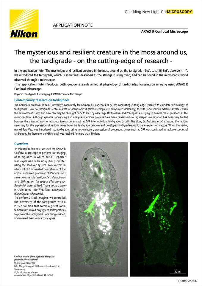
Confocal images of the Hypsibius exemplaris
(Eutardigrada : Parachela)
Vector : pMiUBB-mEGFP
(a) : Merged image of TD (Transmission detector) and fluorescence
(b) : Fluorescence image
Objective lens : Apo LWD 40x WI λS DIC N2
Fig. 1. Confocal images of the Hypsibius exemplaris (Eutardigrada : Parachela)
(a) pMiUBB-mEGFP, (b) pRvUBC-mEGFP
Z Range : 33 µm Z step : 1 µm
Objective lens : Apo LWD 40x WI λS DIC N2
The AX/AX R Confocal Microscope is equipped with a transmission detector (TD) and can simultaneously acquire transmission images using laser scanning and fluorescence, making it easy to identify where fluorescence is occurring within the body. Live imaging using fluorescence and TD confirmed that ubiquitin promoter to work in a part of the muscle (Figures 1 and 2). In both TD and fluorescence, the autofluorescence of chlorella (tardigrade’s food) can be seen in the midgut.
Next, in order to take images at high speed and with less phototoxicity, we utilized the resonant scanning mode. With AX R’s Resonant scanning, it was possible to achieve image quality similar to a Galvano scanning image by averaging about 4 times and processing with Denoise.ai (Figure 2). In addition, by limiting the averaging time to 4 times, it is possible to capture images approximately 3.7 times faster than Galvano scanning. When performing time-lapse imaging, the use of Resonant scanning is useful because it can increase fps.
The fusion of cutting-edge research techniques and imaging instrumentation will contribute to the elucidation of the mysterious ecology of tardigrades, which is still unknown.
Fig. 2. Comparison of confocal images and image processing of the Hypsibius exemplaris (Eutardigrada : Parachela)
(a) Scan mode : Galvano
Averaging time : 1X
(b) Scan mode : Resonant
Averaging time : 4X
Denoise.ai processing
Vector: pMiUBB-mEGFP
Z Range : 33 µm
Z step : 1 µm
Objective lens : Apo LWD 40x WI λS DIC N2
Movies
References
In vivo expression vector derived from anhydrobiotic tardigrade genome enables live imaging in Eutardigrada
Proc Natl Acad Sci U S A . 2023 Jan 31;120(5):e2216739120
Sae Tanaka, Kazuhiro Aoki, and Kazuharu Arakawa
https://doi.org/10.1073/pnas.2216739120
Acknowledgments
We would like to express our deepest gratitude to Dr. Kazuharu Arakawa of the Institute for Advanced Biosciences, Keio University, and Dr. Sae Tanaka of the Exploratory Research Center on Life and Living System, National Institutes of Natural Sciences, for providing samples and providing guidance regarding research in the preparation of this application note.
Dr. Arakawa’s Laboratory URL : https://bioinformatician.org/glab/
Product Information
Supports high-speed, high-resolution, large field-of-view confocal imaging with low phototoxicity to living cells and low photobleaching.
- High Speed : Up to 720 frames per second (Resonant 2048 x 16 pixels)
- High resolution : Up to 8K (Galvano) / 2K (Resonant)
- High throughput : Ultra-wide field of view of 25 mm
