- en Change Region
- Global Site
- Home
- Products
- Digital Microscopes
- ECLIPSE Ui
Interviews
Customer Interview A step up in digital pathology using innovative digital microscopy.
Kitasato University Kitasato Institute Hospital
Director, Department of Pathology
Dr. Ichiro Maeda
Currently there are various treatment methods under development that could provide optimal medical care in response to the pathological attributes of each patient. Therefore, the role required of pathological diagnosis, which is indispensable for the planning of treatment policies and strategies, is becoming more important than ever before. However, the shortage of trained pathologists who assume this role is a common issue, not only in Japan, but also worldwide. To solve this problem, digital pathology has been attracting attention. In this article, we asked Dr. Ichiro Maeda, Director of the Department of Pathology, Kitasato University Kitasato Institute Hospital, for his impressions of using the digital image display optical microscope "ECLIPSE Ui".
*Dr. Maeda has provided feedback to Nikon on this microscope’s features and clinical usefulness based on his own personal experience.
What are your impressions of using the ECLIPSE Ui?
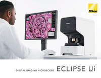
Dr. Maeda: I thought the ECLIPSE Ui would be a very advanced design that would be a little unfamiliar to the unglamorous pathology department. I like the slimmed-down configuration because there are only two buttons, Power and Eject, on its front. It is also stylish, which makes my desk feel a bit sophisticated. The ECLIPSE Ui is compact in size, about the same as a standard optical microscope,so this digital unit fits into a small space to replace the optical one comfortably.
Samples are easy to place on to the stage. There is no need to hold a sample with one hand and to open and set the slide clip with the other hand, as with an optical microscope. Just place the sample and press a button, and then the microscope is set to image with the 4x objective lens. I also think the operability is very well thought out. There are a wide range of shortcut keys for various functions, and if you learn the key arrangement, you can continue to observe samples without looking away from their images on screen. This allows you to observe in a concentrated manner.
In what situations do you use it?
Dr. Maeda: Pathology whole slide imaging diagnostic assist devices (WSI scanners) are classified as a controlled medical device (Class II) that can be used by medical doctors to assist in diagnosis. They allow images to be viewed without having a prepared sample at hand, but it poses a high risk to patients. The ECLIPSE Ui is classified as a digital image display optical microscope that can be used for observation at the discretion of a doctor, even though it is a general medical device (Class I). This is because it displays live images of prepared samples at hand. This medical digital microscope is unprecedented for its real-time display of digital images. Observation can be made as soon as a sample is prepared, without having to capture and check the images in advance. Since the prepared sample is at hand, it is possible to retrieve target cases using the sample’s barcode on a computer of the pathology department system. In other words, the introduction of a WSI system requires high costs, such as purchasing a WSI scanner, securing storage, changing and implementing the system, as well as human resources who are familiar with it. In contrast, with the ECLIPSE Ui in place, the current workflow and system can be kept intact. This digital medical device alone could serve well, as I see it, without hassle.
What samples do you observe?
Dr. Maeda: We observe all the samples that come up in our regular practice. Eosinophil counting has become mandatory for the diagnosis of specified intractable diseases, such as eosinophilic sinusitis and eosinophilic gastroenteritis, and we check more often for eosinophils. Personally, I find it a bit difficult to check for eosinophils with conventional WSI scanners. It seems hard to accurately identify Helicobacter pylori (H. pylori) in gastric biopsy specimens. The ECLIPSE Ui has an objective lens magnification of up to 40x and supports the z axis and depth observation. In addition, you can switch to high-resolution mode (HQ mode) with the touch of a button, so you can observe specific areas in high resolution. For this reason, I was surprised to see eosinophils and H. pylori clearly visible in the digital images of HE-stained samples. Furthermore, the digital zoom function may be used together to observe samples necessary for magnifications of 40x-60x.
For histopathological diagnosis, paraffin-embedded tissue slices are HE-stained or immunohistochemically stained for viewing. The sample size is in the range of 1-30 mm, in which the sample must be observed overall. In order to do this, we scroll sequentially to check for abnormalities. What surprises me is that the images are digital, but there is almost no image distortion or afterimage, even during the scrolling. As with optical microscopes, the images look very natural.
Besides, WSI scanners, classified as pathology whole slide imaging diagnostic assist devices (Class II), have limitations on the types of samples. The ECLIPSE Ui that falls in the category of "Digital Image Display Optical Microscope", a general medical device (Class I), has the advantage that samples prepared on slides are not limited by their types. Even in the cases of intraoperative rapid observation and pathological cytology, this device provides a definitive observation on the spot. This practice may be included in the medical fees. I also feel that the new system can be used in a wide range of applications for routine pathological observation tasks.
What are the advantages of digital image display optical microscopes over optical microscopes?
Dr. Maeda: On optical microscopes, you look through the eyepieces. When you observe over a long time, you could get a stiff neck and feel discomfort in your shoulders and back.
Using the ECLIPSE Ui, I look at the monitor screen and settle into a comfortable position. Unfortunately, with the optical microscope, it is awkward to take off my reading glasses before looking into the eyepieces, put them on after observation, and take them off again to get back to the microscope. It was a bit of hassle. Compared to this, I find it user-friendly when using the monitor screen to continue monitoring and making a observation with or without the reading glasses, and then write a report. The device is equipped with the Trace Display function in order to have an overview of the entire prepared sample, which helps reduce the risk of oversight.
What role do you expect digital microscopes to have in the future?
Dr. Maeda: There are two full-time pathologists in our department, and we welcome part-time pathologists who are department heads from other hospitals so that we can double-check almost all cases. About 90% of pathological diagnoses can be made immediately by experienced pathologists, whereas for 10% or so of them we consult with the part-time doctors and we ask external pathologists for another 10% of the latter. The Department of Pathology is the only department that mainly performs final diagnoses, in other words, a department that involves 100% diagnosis. For this reason, it is common and very important to prepare a letter and a specimen for consultation with a pathologist who specializes in the field in question.
Currently, about 20% of 1000 or so medical institutions with diagnostic pathology departments do not have any full-time pathologists. Even in facilities that are accredited and registered by the Japanese Society of Pathology, more than 50% of hospitals with 200 or more beds are without a pathologist or with a "single pathologist" who we would call. This means that more than 70% of hospitals are without a pathologist or with only 1 or 2 pathologists. In such a dire situation, it is difficult to conduct routine consultations and it is ideal to have systems for double-checking.
On the other hand, AI technology is also being developed in the world of pathology. Although this technology does not serve to make final diagnoses, I think it is effective as a support for pathologists, such as double-checking. In addition, it can be said that the Internet has now come to stay as an infrastructure that supports society, and that real-time remote diagnosis is in place.
Under these circumstances, I believe that digitizing microscopic images is essential to create a sustainable environment for pathological diagnosis. With the ability to digitize pathological sample images, we have the potential to easily overcome the above problems. Until now, WSI scanners have been mainstream for pathological diagnosis using digital images. With the addition of a "digital image display optical microscope" for real-time observation, I expect that the digitization of pathology will advance further.
The ECLIPSE Ui is equipped with an alignment mode, which automatically aligns two different types of samples and displays them side by side on the screen. In this mode, the images can also be repositioned, even if rotated. Going digital gives us such an advantage too.
- Home
- Products
- Digital Microscopes
- ECLIPSE Ui


